Advances in Animal and Veterinary Sciences
Research Article
Advances in Animal and Veterinary Sciences. 2 (3S): 23 – 27Special Issue–3 (Approaches in Diagnosis and Management of Diseases of Livestock and Poultry)
Immunohistochemical Detection of p53 Tumor Suppressor Protein in Round Cell Tumors of Dogs in Grenada, West Indies
Keshaw Prasad Tiwari1, Muhammad Iqbal Bhaiyat1, Alfred Chikweto1, Allison Inga1, Ravindra Nath Sharma1*, RVS Pawaiya2
- Pathobiology Academic Program, School of Veterinary Medicine, St. George’s University, Grenada, West Indies
- Central Institute for Research on Goats, Makhdoom, P.O. Farah–281122, Mathura, Uttar Pradesh, India
*Corresponding author:rsharma@sgu.edu
ARTICLE CITATION:
Tiwari KP, Bhaiyat MI, Chikweto A, Inga A, Sharma RN and Pawaiya RVS (2014) Immunohistochemical detection of p53 tumor suppressor protein in round cell tumors of dogs in grenada, West Indies. Adv. Anim. Vet. Sci. 2 (3S): 23 – 27.
Received: 2014–01–21, Revised: 2014–02–23, Accepted: 2014–02–24
The electronic version of this article is the complete one and can be found online at
(
http://dx.doi.org/10.14737/journal.aavs/2014/2.3s.23.27
)
which permits unrestricted use, distribution, and reproduction in any medium, provided the original work is properly cited
ABSTRACT
A study was undertaken on 40 round cell tumors in dogs namely, transmissible venereal tumor (TVT), 19 cases; mast cell tumor (MCT), 7 cases; canine cutaneous histiocytoma (CCH), 14 cases. These tumor cases were selected from biopsy and necropsy submissions for the period 2001 to 2011 on dogs residing in Grenada. The objective of this study was to evaluate the expression pattern of the tumor suppressor protein p53 in these tumors. Nuclear expression of p53 was detected as moderate to strong immuno–reactivity in most of the 3 tumors under study. The percentage of immuno–marked cells was 36.8, 50.0, and 57.1 for TVT, CCH and MCT, respectively. Intensity of p53 immunostaining was proportional to biological behavior of the tumors. Canine TVT in metastatic sites showed significantly (P=0.009) higher immune–reactivity compared to TVT in primary site. Strongly positive p53 expression was seen consistently in TVT at extragenital sites indicating a higher degree of malignant behavior of this tumor. This suggests that p53 expression could be a useful indicator of the malignant potential and prognosis of canine TVT.
INTRODUCTION
The p53 (also known as protein 53 or tumor protein 53) is a tumor suppressor protein that plays a central role in the maintenance of genomic integrity. p53 has been described as "the guardian of the genome", referring to its role in conserving stability of the cell genome by preventing mutation (Lane, 1992; Strachan and Read, 2003). The name is due to its molecular mass which is 53 kilo Dalton (kDa). It regulates the cell cycle and, thus, functions as a tumor suppressor that is involved in preventing cancer.
The p53 is one of the most important of the tumor suppressor genes involved in the development of neoplasia and the most frequently mutated gene in human cancers (Setoguchi et al., 2001; Soussi and Beround, 2001; Pawaiya and Ramkumar, 2007; Vousden and Lane, 2007). Known biological functions of p53 are transcriptional activation of genes involved in cell cycle control, DNA repair and metabolism, genomic plasticity, senescence, angiogenesis, programmed cell death, cellular differentiation, and immune response, including transcriptional, posttranscriptional and posttranslational roles (Levine, 1997; Hollstein and Hainaut, 2010). p53 protein is quickly eliminated due to its short half–life (about 20 minutes). In contrast, the mutant p53 protein has a half–life of several hours and may be detected by immunohistochemistry (Prokocimer and Rotter, 1994). Correlation between immunohistochemical detection of p53 protein and mutations in p53 gene has been described (Davidoff et al., 1991). These mutations often lead to production of an altered p53 protein that binds to and inactivates the normal p53 protein, thereby promoting tumorigenesis. Thus, immunohistochemical detection of p53 protein is equated to the detection of the mutant p53 protein or otherwise stabilized abnormal p53 protein, rather than due to overexpression of normal p53 (Ginn et al., 2000).
Genetic instability is the one of the most common features of malignant neoplasms. P53 can arrest cell division, freezing the cell at the G1 checkpoint of the cellular cycle, thus the cell is unable to reproduce and its damaged genome is safely isolated. p53 can also initiate a more permanent solution namely programmed cell death or apoptosis if it fails to repair the damaged DNA (Hanahan and Weinberg, 2011; Ozer et al., 2012).
Canine round cell tumors (RCTs) are composed of cells with a round morphology. They include transmissible venereal tumor (TVT), histiocytoma, lymphoma, melanoma, plasmacytoma, and mast cell tumor and neuroendocrine cell tumor (Sandusky et al., 1987). It is difficult to distinguish TVT from other round cell tumors especially when it occurs in extra genital locations.
The aim of present study was to immunohistochemically detect and determine the percentage of cells overexpressing p53 oncoprotein in mast cell tumor, cutaneous histiocytoma and canine transmissible venereal tumor in dogs in Grenada.
MATERIALS AND METHODS
Tumors
Retrospective (archival) canine biopsy and necropsy tumor tissue specimens were obtained from the Veterinary Pathology Laboratory School of Veterinary Medicine, St. George’s University, Grenada collected between 2001 and 2011. Based on histological features, tumor selected and included in the study were 19 TVT, 14 cutaneous histiocytoma and 7 mast cell tumors. No attempt was made to select cases on basis of criteria such as sex, age, or breed of the dog.
Slide Pre–Treatment (Adhesive Coating)
Before taking paraffinized tissue sections glass slides were coated with 3–Aminopropyltriethoxysilane (Sigma–Aldrich, Product No. A3648), following the procedure of Herrington and McGee (1992) and Pawaiya and Ramkumar (2007).
Sectioning and Histological Examination
Histologic sections were cut at 4µm thick from paraffin–embedded blocks of each of the tumors and stained with hematoxyline and eosin (HE). Duplicate, 4µm thick paraffin sections were also taken on adhesive coated slides for immunohistochemical studies. Histologically, each tumor was classified and identified as mast cell tumor (Grade I, II and III), transmissible venereal tumor and cutaneous histiocytoma.
Immunohistochemistry
Immunodetection System for Specific Antibodies
All sections were stained by the Peroxidase immunostaining method using a Mouse ExtrAvidin Peroxidase staining kit (Cat. No. EXTRA–2; Sigma–Aldrich, USA) with mouse Monoclonal anti–p53 oncoprotien serum as the primary antibody (Clone No. BP53–12; Product No. 5813; Sigma–Aldrich, USA) and biotinylated anti–mouse Ig G serum as secondary antibody. Antibody localization was determined using 3–amino–9–ethylcarbazol (AEC) substrate as the chromogen.
Antigen Retrieval
Antigen unmasking on paraffin sections was carried out following the method of Cattoretti et al. (1993) with modifications as described by Pawaiya and Ramkumar (2007).
Immunohistochemical Staining
Immunostaining protocols supplied by the manufacturers along with the antibodies and immuno–detection kits were followed with suitable modifications as per Pawaiya and Ramkumar (2007).
Paraffin–embedded tissue sections were deparaffinized and brought to water by sequential immersion in xylene and graded ethanol concentrations. Sections were then rinsed in distilled water and immersed in antigen retrieval solution (0.01M Citrate buffer, pH 6.0) heated to boil in a microwave oven 3 times for 5 min each and cooled to the room temperature by leaving as such. Endogenous peroxidase activity was quenched by immersion in 3% hydrogen peroxide in absolute methanol for 20 minutes at room temperature and then tissue sections were incubated with 5% normal goat serum (Product No. G9023; Sigma–Aldrich, USA) for 30 min. The primary antibody was diluted (1:100) in PBS with 1% Bovine serum albumin (Product No. A7030; Sigma–Aldrich, USA) and 0.1% sodium azide (as preservative) and applied adequately (100–150 µl) to cover the moist tissue sections and incubated for 1 hr in a humidified chamber at room temperature (RT). Biotinylated goat anti–mouse IgG antibody (1:15 dilution) was used as secondary antibody with which the sections were incubated for 30 min, followed by incubation in ExtrAvidin peroxidase (1:15) in humidified chamber at RT for 30 min. Working solution of AEC chromogen substrate was applied on moist sections and incubated for 10–15 min or till the brick–red color appeared whichever was earlier. The reaction was terminated by rinsing with distilled water. AEC stained sections were counterstained with Mayer's haematoxylin (Product No. MHS–16; Sigma–Aldrich, USA) and mounted with aqueous mounting medium CC Mount (Product No. C9368; Sigma–Aldrich, USA).
The selected tumor sections for IHC staining, in which the primary antibody was replaced by normal goat serum, were used as negative controls. Each slide acted as an internal positive control, because those cells in the basal layer of the epidermis are normally dividing. All the procedural steps were interceded by washing thrice (5 min each) in phosphate buffered saline (pH 7.4).
Stained sections were examined under light microscope. Ten fields per tissue section covering highly cellular areas were counted under 40x magnification objective using image analysis software (NIS–Elements AR, Nikon). Brick–red staining within the nuclei of tumor cells was considered to be positive for p53 oncoprotein overexpression. The results were recorded as positive or negative immunolabelled p53 for tumor cells. For each slide, both positively and negatively stained tumor cells were counted and the overall percentage of positively stained tumor cells was calculated.
Statistical Analysis
The data obtained were analyzed by the statistical methods (Fisher’s exact test) using a graph pad statistical software (http://www.graphpad.com/quickcalcs/contingency2).
RESULTS
Based on histological features, tumors were diagnosed as TVT, CCH and MCT. Immunohistochemistry revealed the presence of p53 positively stained nuclei in the neoplastic cells in these tumors (Figure 1, 2&3). Moderate to strong p53–positive nuclear immunoreactiviy was noticed in the TVT (Figure 1A, B) and CCH (Figure 2A, B) with more cells showing immunopositivity in the former (39.45±22.82) compared to CCH (19.96±5.39). Canine Mast cell tumors though exhibited almost similar count of immunopositively stained cells (20.09±5.40) like CCH, the intensity of immunostaining was moderate to weak (Figure 3A, B).
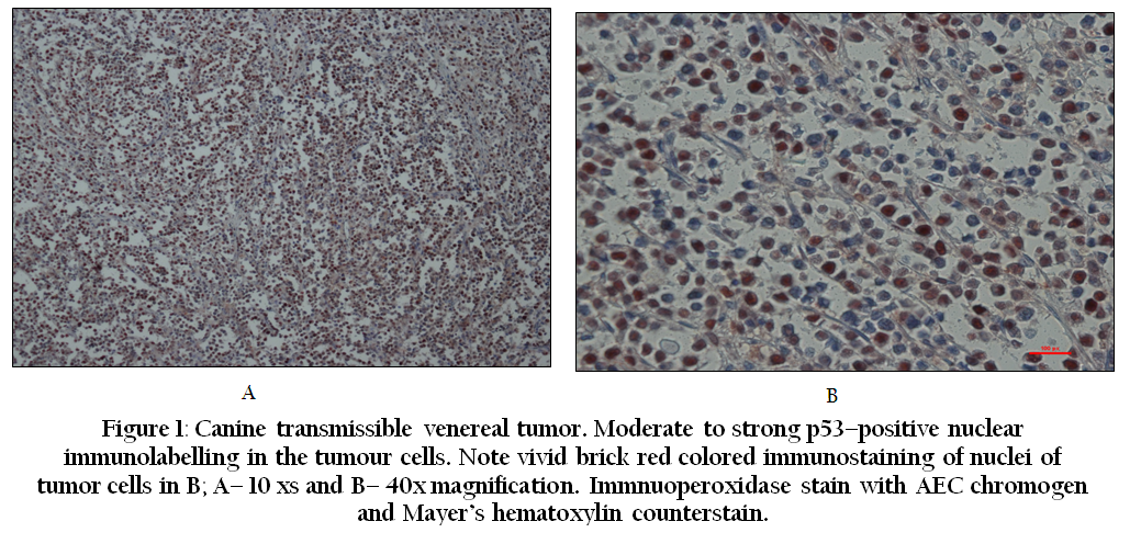
Figure 1: Canine transmissible venereal tumor. Moderate to strong p53–positive nuclear immunolabelling in the tumour cells

Figure 2: Canine cutaneous histiocytoma. Moderate p53–positive nuclear immunolabelling in the tumour cells
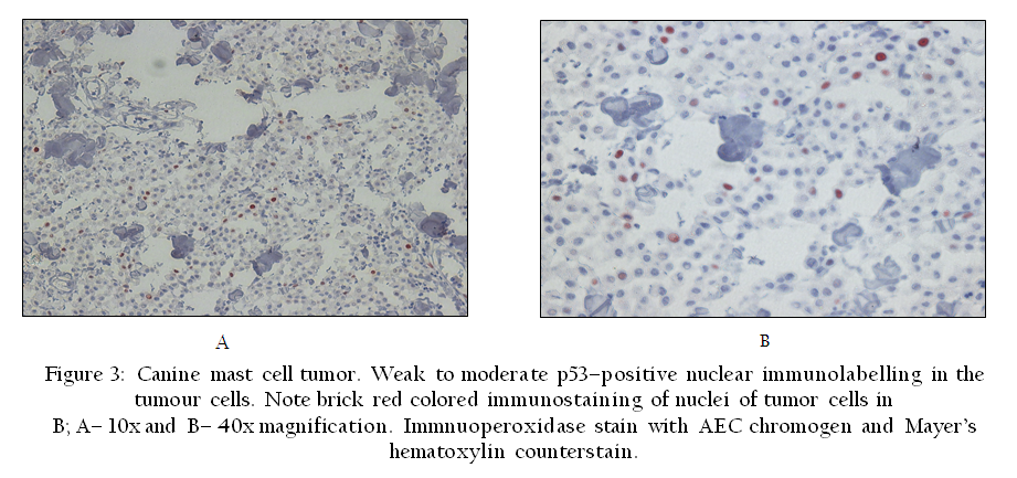
Figure 3: Canine mast cell tumor. Weak to moderate p53–positive nuclear immunolabelling in the tumour cells
Individually, 7 (36.84%) of 19 TVT specimens, 7 (50%) of 14 CCH specimens and 4 (57.14%) of 7 MCT specimens showed p53–positive immunolabelling (Table 1). Canine TVT in metastatic sites (liver, spleen, lymph nodes, mammary gland, maxilla, eye orbit and skin) showed significantly (P = 0.0090) more p53–positive immunolabelling compared to TVT located on the primary natural sites. Overall, out of the 40 round cell tumors included in the study 45% (18/40) revealed p53–positive immunoreactivty (Table 1).
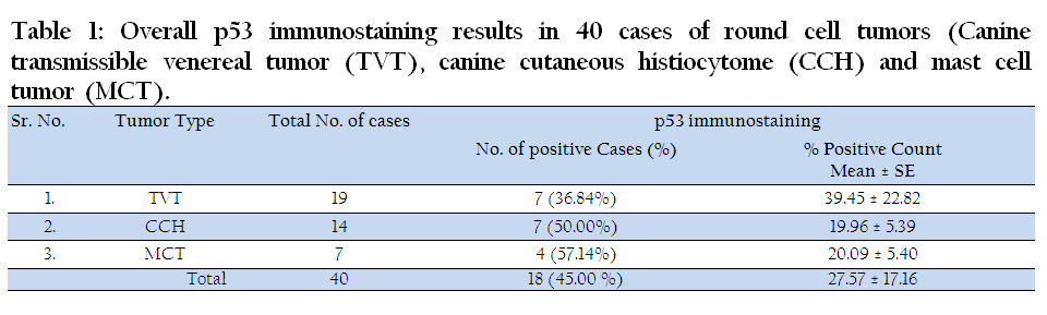
Table 1: Overall p53 immunostaining results in 40 cases of round cell tumors (Canine transmissible venereal tumor (TVT), canine cutaneous histiocytome (CCH) and mast cell tumor (MCT)
DISCUSSION AND CONCLUSIONS
The p53 immunohistochemistry indicated moderate to strong p53–positive nuclear immunolabelling in 7 cases of TVT, while 12 of these tumors did not show any immunostaining. The reason for the negative or weak immunoreactivity of p53 in these specimens may be due to either inactivation of p53 gene in some of the TVT cases examined or inadequate reaction between antihuman p53 antibodies with canine tissues.
The activation of this protein occurs in response to stress or damage to cellular DNA, causing cell cycle arrest and the induction of senescence or apoptosis. More than 10 mutations in the P53 gene have been described in canine neoplasias (Oren, 1999; Setoguchi et al., 2001), including cases of TVT (Choi and Kim, 2002; Sánchez–Servín et al. 2009; Stockmann et al. 2011). Moro et al. (2010) found more cells that expressed p53 protein in transplanted TVT in the regression phase compared with naturally occurring TVT. These findings suggested that there may be functional abnormalities in the p53 gene and its transcription in these tumors (Stockmann et al. 2011).
Overexpression of p53 protein is a good, but not unique, indicator of mutation in p53 gene (Vogelstein and Kinzler, 1992). Other investigators have indicated that p53 detected by immunohistochemical evaluation was not always mutant (Roels et al., 2001). In human tumors, positive immunohistochemical staining for p53 is often accepted as evidence of an underlying p53 genetic abnormality and as there is limited DNA sequence data available to verify this conclusion in dogs, the potential for detection of the normal p53 protein in this species merits consideration (Wolf et al., 1997). Increased levels of p53 oncoprotein have been detected immunohistochemically in canine mammary tumors, mast cell tumor, osteosarcoma, Sertoli–cell tumor, seminoma, cutaneous histiocytoma, squamous cell carcinoma, perianal gland adenocarcinoma, hemangiopericytoma, apocrine gland, intestinal and nasal adenocarcinomas, and colorectal tumors (Sagartz et al., 1996; Gamblin et al., 1997; Wolf et al., 1997; McEntee and Brenneman, 1999; Ginn et al., 2000; Inoue and Wada, 2000; Jaffe et al., 2000; Setoguchi et al., 2001).
The intensity and proportion of expression of p53 protein have been considered a potential factor for prognosis in several tumors. A positive correlation was verified between p53 overexpression and prognosis in canine osseous tumors (Sagartz et al., 1996), astrocytomas (Stoica et al., 2004), and mammary tumors (Lee et al., 2004), but not in gastrointestinal mastocytomas (Ozaki et al., 2002), colorectal epithelial tumors (Wolf et al., 1997), and canine and feline melanomas (Roels et al., 2001).
It has been observed that canine TVT of natural occurrence as well as those TVT resistant to chemotherapy showed the weak reactivity to p53 protein (Moro et al., 2010), suggesting that the p53 gene is not involved in this type of tumor, at least alone, with resistance to chemotherapy (Harris and Hollstein, 1993). In present study p53–negative immunostaining for TVT cases found on natural sites (penis, prepuce, vulva, and vagina) are consistent with the observations of Moro et al. (2010). On the other hand, strong p53–positive nuclear immunolabelling was observed in TVT metastasized to other visceral organs (such as liver, spleen, mammary glands, lymph nodes, eye orbit etc), indicating the higher degree of malignant behavior of the tumors. In present study strong p53–positive nuclear immunoreactivity was observed in 3/7 TVT metastasized to other visceral organs which is consistent with the findings of earlier workers (Jaffe et al. 2000; Muto et al., 2000; Sagartz et al., 1996). These findings suggested that p53 immunolabelling could be useful in determining the degree of malignant behavior, and thus the prognosis of canine transmissible venereal tumors.
Conclusively, malignant TVT exhibited the highest percentage of immunoreactivity with p53, and a significant difference was observed with non–metastatic TVT. Non–metastatic TVT located in natural sites showed either negative or low reactivity to p53 protein.
Canine cutaneous histiocytoma (CCH) is another common round cell tumor which has almost histological features similar to TVT. Out of 14 cases of the CCH, 7 (50%) showed positive immunoreactivity to p53. Mostly the tumor cells revealed low to moderate staining intensity, ranging from 13.86% to 29.4%. Overexpression of p53 in CCH has been observed by several workers, with varied degree and intensity of immunolabelling varied significantly (Gamblin et al., 1997; Pawaiya, 2005). Pawaiya et al. (2008) recorded strong immunoreactivity to p53 in a rare case of cutaneous histiocytoma in a buffalo. The observation of moderate overexpression of p53 in half of the CCH cases in the present study indicates the benign nature of the tumor as compared to the higher immunostaining intensity observed in malignant TVT.
Among the 7 cases of mast cell tumors, one was histopathologically diagnosed as a grade–II MCT while the other 6 were grade–I MCTs. Four of the 6 grade I MCT showed positive immunostaining for p53. Our findings of 57% positivity of MCT for p53 overexpression exceeded those reported by Ginn et al. (2000) and Gamblin et al. (1997) who reported positivity of 47% (25/53) and 25% (4/16), respectively.
In this study, the percentage of tumor cells exhibiting p53–positive nuclear immunolabelling for MCT ranged from 12.68% to 25.56%. Jaffe et al (2000) observed a significantly greater percentage of mast cells staining for p53 in grade III tumors than grade I and II tumors, but no difference between grade I and II tumors. They suggested that the overlap of the ranges of the percentage of p53–stained cells made this criterion unreliable for discriminating between histologic grades. They also noticed a high percentage of cells staining for p53 in some grade I MCTs, whereas a very low percentage in some grade III tumors. Previous subjective clinical observations have suggested that some grade I mast cell tumors behave malignantly while some grade III mast cell tumors behave benignly (Bostock, 1973; Patnaik et al. 1984). Our findings show that immunohistochemical staining for p53 offers no advantage over histopathologic grade with regard to the association with survival time or time to recurrence (prognosis) which concurs with the observation of Ginn et al. (2000).
ACKNOWLEDGEMENTS
The authors would like to thank Mrs. Carla Richards, Mrs. Vanessa Mathew for their preparation of histological section and technical potential for IHC staining and Mr. Ray Samuel for technical support. The facilities and equipment of the diagnostic Veterinary pathology laboratory of St. George’s University, School of Veterinary Medicine were used to conduct this study.
REFERENCES
Bostock D (1973). The prognosis following surgical removal of mastocytomas in dogs. J Small Anim. Prac. 14: 27–40.
Cattoretti G, Pileri S, Parravicini C, Becker MHG, Poggi S, Bifulco C, Key G, D'Amato L, Sabattini E, Feudale E, Reynolds F, Gerdes J and Rilke F (1993). Antigen unmasking on formalin–fixed, paraffin–embedded tissue sections. J Pathol. 171:83–98.
http://dx.doi.org/10.1002/path.1711710205
PMid:7506771
Choi YK and Kim CJ (2002). Sequence analysis of canine LINE–1 elements and p53 gene in canine transmissible venereal tumor. J Vet. Sci. 3: 285–292.
PMid:12825561
Davidoff AM, Humphrey PA, Iglehart JD and Marks JR (1991). Genetic basis for p53 overexpression in human breast cancer. Proc Natl Acad. Sci. 88: 5006–5010.
http://dx.doi.org/10.1073/pnas.88.11.5006
PMid:2052583 PMCid:PMC51796
Gamblin RM, Sagartz JE and Couto CG (1997). Overexpression of p53 tumor suppressor protein in spontaneously arising neoplasms of dogs. Am J Vet. Res. 58: 857–863.
PMid:9256970
Ginn PE, Fox LE, Brower JC, Gaskin A, Kurzman ID and Kubilis PS (2000). Immunohistochemical detection of p53 tumor–suppressor protein is a poor indicator of prognosis for canine cutaneous mast cell tumors. Vet. Pathol. 37: 33–39.
http://dx.doi.org/10.1354/vp.37-1-33
PMid:10643978
Graph pad statistical software. Available at URL: http://www.graphpad.com/quickcalcs/contingency2). Accessed on 08 December, 2013.
Hanahan D and Weinberg RA (2011). Hallmarks of Cancer: The Next Generation. Cell 144: 646–676.
http://dx.doi.org/10.1016/j.cell.2011.02.013
PMid:21376230
Harris C and Hollstein M (1993). Clinical implications of the p53 tumor suppressor gene. N Engl J Med 329: 1318–1327.
http://dx.doi.org/10.1056/NEJM199310283291807
PMid:8413413
Herrington CS and McGee JO'D (1992). Diagnostic Molecular Pathology: A Practical Approach. Oxford University Press, Oxford 1: 80.
Hollstein M and Hainaut P (2010). Massively regulated genes: the example of TP53. J Pathol. 220:164–173.
PMid:19918835
Inoue M and Wada N (2000). Immunohistochemical detection of p53 and p21 proteins in canine testicular tumors. Vet. Rec. 146:370–372.
http://dx.doi.org/10.1136/vr.146.13.370
PMid:10803982
Jaffe MH, Hosgood G, Taylor HW, Kerwin SC, Hedlund CS, Lopez MK, Davidson JR, Miller DM and Paranjpe M (2000). Immunohistochemical and clinical evaluation of p53 in canine cutaneous mast cell tumors. Vet. Pathol. 37: 40–46.
http://dx.doi.org/10.1354/vp.37-1-40
PMid:10643979
Lane DP (1992). Cancer p53, guardian of the genome. Nature 358: 15–16.
http://dx.doi.org/10.1038/358015a0
PMid:1614522
Lee CH, Kim, WH, Lim JH, Kang MS Kim DY and Kweon OK (2004). Mutation and over expression of p53 as a prognostic factor in canine mammary tumors. J vet. Sci. 5:63–69.
http://dx.doi.org/10.1292/jvms.66.63
PMid:15028887
Levine AJ (1997). p53, the cellular gatekeeper for growth and division. Cell 88(3):323–31.
http://dx.doi.org/10.1016/S0092-8674(00)81871-1
McEntee MF and Brenneman KA (1999). Dysregulation of beta–catenin is common in canine sporadic colorectal tumors. Vet. Pathol. 36: 228–236.
http://dx.doi.org/10.1354/vp.36-3-228
PMid:10332831
Moro JV, Tinucci–Costa M,Silveira ACT, Gerardi DG and Alessi AC (2010). Reactivity of p53 protein in canine transmissible venereal tumor. Arquivo Brasileiro de Medicina Veterinária e Zootecnia 62: 318–323.
http://dx.doi.org/10.1590/S0102-09352010000200011
Muto T, Wakui S, Takahashi H, Maekawa S, Masaoka T, Ushigome S and Furusato M (2000). p53 gene mutations occurring in spontaneous benign and malignant mammary tumors of the dog. Vet. Pathol. 37(3):248–53.
http://dx.doi.org/10.1354/vp.37-3-248
PMid:10810989
Oren M. (1999). Regulation of the p53 tumor suppressor protein. J Biol. Chem. 274: 36031–36034.
http://dx.doi.org/10.1074/jbc.274.51.36031
PMid:10593882
Ozaki K, Yamagami T.,Nomura K. and Narama I (2002). Mast cell tumours of the gastrointestinal tract in 39 dogs. Vet. Pathol. 39: 557–564.
http://dx.doi.org/10.1354/vp.39-5-557
PMid:12243465
Ozer H, Yenicesu G, Arici S, Cetin M, Tuncer E and Cetin A (2012). Immunohistochemistry with apoptotic–antiapoptotic proteins (p53, p21, bax, bcl–2), c–kit, telomerase, and metallothionein as a diagnostic aid in benign, borderline, and malignant serous and mucinous ovarian tumors. Diagn. Pathol. 7: 124. doi: 10.1186/1746–1596–7–124.
http://dx.doi.org/10.1186/1746-1596-7-124
Patnaik AK, Ehler WJ, and MacEwen EG (1984). Canine cutaneous mast cell tumor: morphologic grading and survival time in 83 dogs. Vet. Pathol. 21:469–474.
PMid:6435301
Pawaiya RVS and Ram Kumar (2007). Ovine pulmonary adenocarcinoma: evaluation of molecular tumour markers. Indian J Vet. Pathol. 31: 99–107.
Pawaiya RVS (2005). Pathology of chemically induced neoplasms and evaluation of molecular markers in diagnosis of animal tumours. Indian J Vet. Pathol. 29 (1): 63.
Pawaiya RVS, Ramkumar and Pawde AM (2008). Immunohistochemical study of a rare case of cutaneous histiocytoma in buffalo. Indian J Vet. Pathol. 32: 251–256.
Prokocimer M and Rotter V (1994). Structure and function of p53 in normal cells and their aberrations in cancer cells: projection on the hematologic cell lineages. Blood 84: 2391–2411.
PMid:7919359
Roels S, Tilmant K and Ducatelle R (2001). p53 expression and apoptosis in melanomas of dogs and cats. Res in Vet. Sci. 70: 19–25.
http://dx.doi.org/10.1053/rvsc.2000.0435
PMid:11170847
Sagartz JE, Bodley WL, Gamblin RM, Couto CG, Tierney LA and Capen CC (1996). p53 tumor suppressor protein overexpression in osteogenic tumors of dogs. Vet. Pathol.33: 213–221.
http://dx.doi.org/10.1177/030098589603300211
PMid:8801715
Sánchez–Servín A, Martínez S, Córdova–Alarcon E and Fajardo R (2009). TP53 Polymorphisms allow for genetic sub–grouping of the canine transmissible venereal tumor. J Vet. Sci. 10: 353–355.
http://dx.doi.org/10.4142/jvs.2009.10.4.353
PMid:19934603 PMCid:PMC2807274
Sandusky GE, Carlton WW and Wightman KA (1987). Diagnostic immunohistochemistry of canine round cell tumors. Vet. Pathol. 24: 495–499.
PMid:3137715
Setoguchi A, akai T, Okuda M, Minehata K, Yazawa M, Ishizaka T, Watari T, Nishimura R, Sasaki N, Hasegawa AT and Sujimoto H (2001). Aberrations of the p53 tumor suppressor gene in various tumors in dogs. Am J Vet. Res. 62: 433–439.
http://dx.doi.org/10.2460/ajvr.2001.62.433
PMid:11277210
Soussi T and Béroud C (2001). Assessing TP53 status in human tumours to evaluate clinical outcome. Nature rev Cancer 1: 233–40.
http://dx.doi.org/10.1038/35106009
PMid:11902578
Stockmann D, Ferrari HF, Andrade AL, Lopes RA, Cardoso TC and Luvizotto MCR (2011). Canine Transmissible Venereal Tumors: Aspects Related to Programmed Cell Death. Braz J Vet. Pathol. 4: 67–75.
Stoica G, Kim HT, Hall DG and Coates JR (2004). Morphology, immunohistochemistry and genetic alterations in dog astrocytomas. Vet.Pathol. 41: 10–19.
http://dx.doi.org/10.1354/vp.41-1-10
PMid:14715963
Strachan T and Read A (2003). Human Molecular Genetics. 3rd edition. Garland Science/Taylor & Francis Group. New York.
Vogelstein B and Kinzler KW (1992). p53 function and dysfunction. Cell 70: 523–526.
http://dx.doi.org/10.1016/0092-8674(92)90421-8
Vousden KH and Lane DP (2007). p53 in health and disease. Nature Rev Mol. Cell. Biol. 8: 275–283.
http://dx.doi.org/10.1038/nrm2147
PMid:17380161
Wolf J, Ginn P, Homer B, Fox L and Kurzman I (1997). Immunohistochemical detection of p53 tumor suppressor gene protein in canine epithelial colorectal tumors. Vet. Pathol. 34: 394–404.
http://dx.doi.org/10.1177/030098589703400503
PMid:9381650


