Advances in Animal and Veterinary Sciences
Research Article
Advances in Animal and Veterinary Sciences. 2 (3S): 8 – 11Special Issue–3 (Approaches in Diagnosis and Management of Diseases of Livestock and Poultry)
Parasitic Encephalomyelitis in Goats due to Aberrant Infestation with Botfly Oestrous ovis Larvae
Nitika Sharma*, Shivasharanappa Nayakwadi, Rajveer Singh Pawaiya, Surender Kumar, Waseem A Tailie, Souvik Paul, Anil Kumar Mishra, Kumaresan Gururaj, Ashok Kumar, Vivek Kumar Gupta, Vinay Chaturvedi
*Corresponding author:nitika4419@gmail.com
ARTICLE CITATION:
Sharma N, Nayakwadi S, Pawaiya RS, Kumar S, Tailie WA, Paul S, Mishra AK, Gururaj K, Kumar A, Gupta VK, Chaturvedi V (2014). Parasitic encephalomyelitis in goats due to aberrant infestation with botfly Oestrous ovis larvae. Adv. Anim. Vet. Sci. 2 (3S): 8 – 11.
Received: 2014–01–21, Revised: 2014–02–18, Accepted: 2014–02–23
The electronic version of this article is the complete one and can be found online at
(
http://dx.doi.org/10.14737/journal.aavs/2014/2.3s.8.11
)
which permits unrestricted use, distribution, and reproduction in any medium, provided the original work is properly cited
ABSTRACT
Nasal myiasis or oestrosis is primarily a disease of sheep (but rarely in goats, camel, deer, dog and man) caused by the infestation of nasal cavities by the larvae of botfly Oestrus ovis. Four adult goats, aged between three and five years were presented with clinical history of onset of episodes of neurological symptoms such as ataxia, staggering gait, convulsions, circling and paddling of legs. The animals were treated but the response to the treatment was very poor. The severity of neurological signs increased with time resulting in recumbency and death of the animals. On necropsy, 19 larvae were recovered from the nasal sinuses and cranial cavity of one animal, 4 from brain of another goat and one larva each from the brain of other two animals. The Oestrus ovis larvae were confirmed based on the presence of posterior ‘D ‘shaped respiratory spiracles. These very rare cases of aberrant infestation of goat brain with Oestrus ovis larvae, their clinical manifestation, pathology and public health significance have been described and discussed in detail.
INTRODUCTION
Oestrosis, also known as nasal myiasis, is a worldwide disease of animals caused by the larvae of botfly, Oestrus ovis (Linne 1761, Diptera: Oestridae), are obligatory parasites of the nasal and sinus cavities of sheep and goats (Gunalan et al., 2011; da Silva et al., 2012; 2013 Hanan, 2013). The parasite primarily infests sheep but rarely infests goats, camel, deer, reindeer, elk, ibex, dog and man (Lapage, 1956; Soulsby, 1982; Das and Bhatia, 1994). Infection occurs due to deposition of eggs by adult fly in the nostrils of the host. Infestation of O. ovis is usually not life threatening, but ocassionally there is bacterial infection that causes encephalitis and death of the host. Secondary infections often lead to mucopurulent discharge, respiratory distress and sneezing fits in the affected animals (Urquhart et al., 2003). Nervous form of O. ovis infestation in sheep and goats is quite infrequent. Rarely, these larvae penetrate into ethmoid bones and reach cranial cavity resulting in abnormal neurological signs such as a high–stepping gait, incoordination and occasional death (Radostits et al., 2007; Shivasharanappa et al., 2011). Neurological signs usually mimic the infection of Coenurus cerebralis because of which this disease is also known as ‘False Gid’. The present documentation reports rare cases of aberrant infestation of goat brain with migrating O. ovis larvae resulting in sudden onset of episodes of neurological signs and fatal parasitic encephalomyelitis.
MATERIALS AND METHODS
History and Clinical Examination
Four adult goats aged between three and five years were presented to the Division of Animal Health of the Institute with a history of onset of episodes of neurological signs such as ataxia, staggering gait, convulsions, circling and paddling of legs. Clinical examination of the animals revealed subnormal body temperature (97– 98oF), pale conjunctival mucus membrane and poor body condition. Vital signs including heart and respiratory rates were normal. Cerebrospinal fluid (CSF) collected from the lumbosacral region was found to be turbid with pleocytosis, elevated level of total proteins (80–90 mg/dl) and reduced glucose level (25–30.8 mg/dl). Differential leukocyte count showed lymphocytosis and neutropenia. Bacteriological examination of CSF and blood was negative. Faecal examination revealed mixed infections of gastro–intestinal nematodes, Moniezia, and Coccidia.
Treatment and Response to Treatment
In all the four animals, treatment was instituted immediately after brief clinical examination as warranted by the acuteness of the condition. Treatment with Sulphadiazine and Trimethoprim combination (Biotrim®) @ 15 mg/Kg BW I/M), Dexamethasone (Dexona®) @ 5 mg/Kg BW I/M and supplementation with vitamin B1, pyridoxine hydrochloride and cyanocobalamine, Neuroxin– 12V®) resulted in slight improvement in the condition. Despite therapy, episodes of neurological signs increased with time, the animal became laterally recumbent and finally succumbed to the disease within three to five days of the initiation of treatment.
Post–Mortem Examination and Sample Collection
All four goats were necropsied thoroughly immediately after death and macroscopic observations including in the nasal, paranasal sinuses and cranial cavities were recorded. The live larvae were carefully removed from the nasal cavities, paranasal sinuses and the brain, and collected in PBS for further morphological examinations grossly as well as under stereoscopic microscope (Soulsby, 1982). The brain was collected in 10% formol saline for histopathological investigation.
RESULTS
On necropsy of one of the four animals, grossly, a total of 19 larvae were recovered, 14 from the paranasal sinuses and 5 from the cranial cavity (Figure 1). Detailed examination of cranial cavity showed severely damaged brain showing necrotic encepahlomalacic and haemorrhagic lesions in the cerebral hemispheres. The left cerebral hemisphere was almost absent and liquefactive as it was completely destroyed by the migrating larvae (Figure 2). The larvae 1 to 3 cm long, creamy, dark brown or blackish in colour, having characteristic dark pigmented respiratory spiracles were seen partially embedded in the necrotic brain parenchyma (Figure 3). Similarly, in the second goat, four larvae were found in the brain parenchyma. In the other two goats, single larva was found in the left cerebral hemisphere. No specific lesions were observed in any other organ systems. In all the four cases, nasal cavity was explored for larvae but no larvae were found.
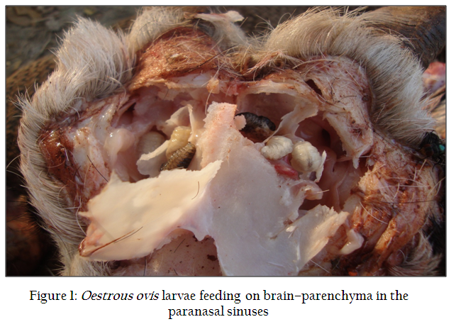
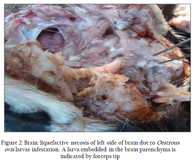
Figure 2: Brain: liquefactive necosis of left side of brain due to Oestrous ovis larvae infestation. A larva embedded in the brain parenchyma is indicated by forceps tip
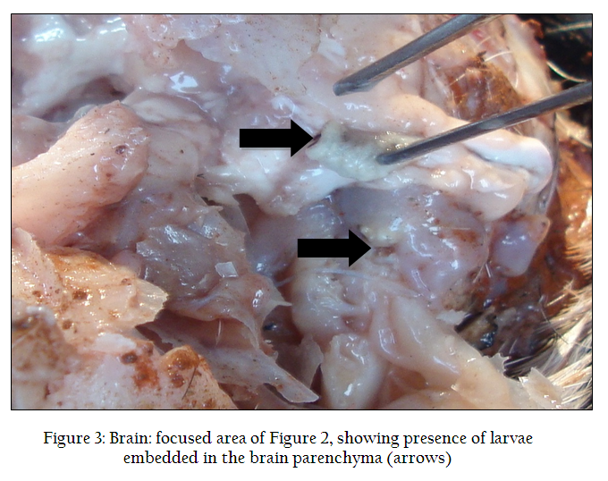
Figure 3: Brain: focused area of Figure 2, showing presence of larvae embedded in the brain parenchyma (arrows)
Histopathological examination of the brain tissue revealed multiple haemorrhagic tracts containing necrotic tissue debris, erythrocytes and inflammatory cells mostly mononuclear cells and a few neutrophils/ eosinophils (Figure 4). Detailed microscopic examination of the recovered larvae revealed transverse bands on dorsal surface of each segment, anterior end was armed with minute paired hooks, posteriorly connected with cephalo–thoraxial skeleton and ventral surface had rows of minute spines. The larvae were confirmed as O. ovis on the basis of ‘D’ shaped, closed, dark brown coloured, deep stigmal plates with radially arranged respiratory holes (Figure 5). Based on the history, clinical signs, laboratory findings, necropsy and pathological findings, these cases were diagnosed as parasitic encephalomyelitis due to aberrant migration of Oestrus ovis larvae with apparent secondary complications.
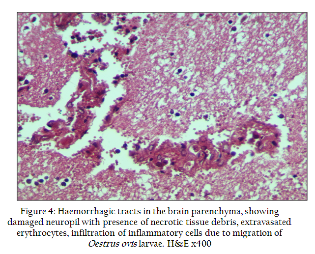
Figure 4: Haemorrhagic tracts in the brain parenchyma, showing damaged neuropil with presence of necrotic tissue debris, extravasated erythrocytes, infiltration of inflammatory cells due to migration of Oestrus ovis larvae. H&E x400
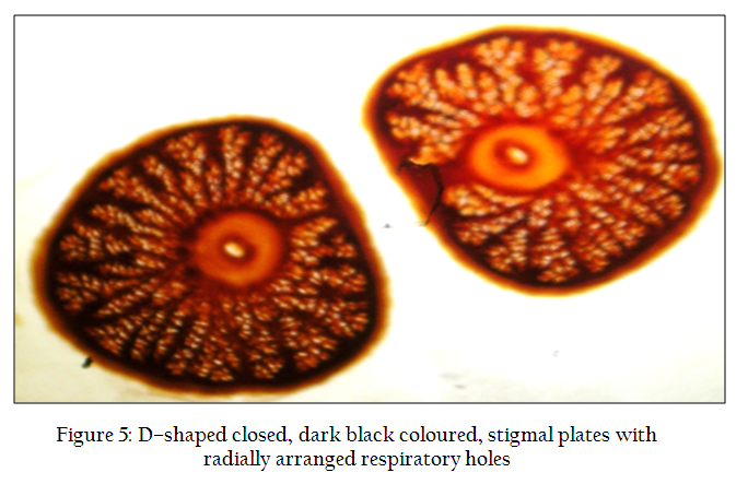
Figure 5: D–shaped closed, dark black coloured, stigmal plates with radially arranged respiratory holes
DISCUSSION
Infection of O. ovis occurs due to deposition of eggs by adult fly in and around the nostrils. The larvae enter the nasal passage where they remain and develop through the second instar. The third instar larvae enter the head sinuses and remain there for 8–10 months. The larvae usually attain full size in late winter or spring and leave the sinuses to pupate. When the third instar larvae reaches the nasal passages, the host sneezes violently thus ejecting the larvae with great force (Soulsby, 1982; Marquardt et al., 2000; Taylor, 2007). The adult larvipositing O. ovis has a short lifespan of just two weeks and is known to possess affinity for its natural host sheep (Godara et al., 2010). Infection rates and larval burdens are always higher in sheep than in goats after either natural or artificial infestation (Duranton et al., 1996; Dorchies et al., 2000; Papadopoulos et al., 2001; 2006).
In the present study, all four cases of goats were reared together with sheep in mixed flocks owned by farmers. In India, the farmers generally rear sheep and goats together (Pathak, 1992). Although O. ovis is primarily a parasite of domestic sheep, other hosts like goats, man, dogs, camels are sometimes infected (Soulsby, 1982; Das and Bhatia, 1994; Abdel–Aziz et al., 2010). In a mixed flock, botfly can also have easy access to infect goats although infection rates and larval burdens of sheep are always significantly higher than goats (Papadopoulos et al., 2001; 2006). Papadopoulos et al. (2006) observed significantly higher infection rates of O. ovis in sheep than goats when compared seroprevalence of O. ovis in mixed flocks. Perhaps, due to close contact of sheep and goats the O. ovis fly deposited its larvae on the face or nostrils of the goat.
During their life cycle, the O. ovis larvae migrate to the dorsal turbinates and frontal sinuses where they remain for few months before they migrate to nostrils and are sneezed out to pupate on the ground (Radostits et al., 2007). Interestingly, it is important to note that one seldom sees more than a single third instar larva in the head sinus. It seems that there is a feed–back mechanism that limits the occupation of a relatively limited space in the natural host sheep (Marquardt et al., 2000). Whether such feed–back mechanism is also at work in the uncharacteristic host goat is not yet known, and probably the absence of this may be the reason that, in the present study, a total of 19 larvae, 14 from paranasal sinuses and 5 from cranial cavity were recovered in one case, while 4 larvae were present in the brain of another case. Normally O. ovis larvae are confined to the nasal cavities and paranasal sinuses of the head. It is very rare and unusual to find such a large number of larvae in the cranial cavity and brain. These larvae might have penetrated into ethmoid bones and reached cranial cavity. These migrating larvae were either feeding on the brain parenchyma and had eaten nearly half of the brain in one case or caused traumatic injury and haemorrhages with consequent secondary bacterial infection leading to liquefactive necrosis and lysis of the half of the brain, as seen grossly and microscopically in the present study. It has been observed that O. ovis larvae are not hematophagus, and usually feed on plasma proteins, antibodies, secreted from the nasal muscosa during inflammatory reaction, mucin, and collagen of the basal membrane which they digest through proteases in their excretory/secretory products (Tabouret et al., 2003; Angulo–Valadez et al., 2010). The episodes of abnormal neurological signs in the affected goat were caused due to the injury to the brain parenchyma by the migration of O. ovis larvae. Microscopic lesions in the brain showed multiple haemorrhagic tracts with infiltration of inflammatory cells and necrosis of parenchyma (neuropil) due to migration of larvae. Mechanical activity of oral hooks and spines as well as and the proteolytic enzymes present in the secretory and excretory products of these larvae were responsible for these changes in the brain (Tabouret et al., 2003). Such irreversible damage to the brain parenchyma resulted in severe form of motor abnormalities such as ataxia, convulsions, paddling of legs and incoordination in the affected goats.
In unnatural hosts, in addition to the nares the flies oviposit on the conjunctiva of the eye, mouth and ear canal. Generally, the infection in unnatural hosts is self–limiting, but a few cases have been recorded in which first, second and third instar larvae have been removed from aberrant locations like eye and head (Marquardt et al., 2000; Abdel–Aziz et al., 2010). Infestation and recovery of O. ovis larvae from the aberrant sites in nonspecific and uncharacteristic hosts (ungulates, man and dog) have been reported from the eyes, eyelids, orbital tissues, mucosal cavity and lips (Hall and Smith, 1993; Abdel–Aziz et al., 2010). However, infestation and recovery of the nasal bots of sheep from the aberrant sites like cranial cavity and brain is very unusual and quite rare with the only one traceable report in a goat (Shivasharanappa et al., 2011). This report documenting four cases of aberrant infestation of goat brain by O. ovis larvae in such a large number contributes significantly to the scarce literature on the subject.
Oestrosis in small ruminants causes widespread morbidity and sometimes mortality which in turn results in heavy economic losses to sheep and goat rearers (Pathak, 1992). Public health significance of O. ovis infestation cannot be undermined as it is associated with human population, when bots are deposited on the aberrant sites causing catarrhal conjunctivitis, corneal opacity, visual impairment (ophthalmo–myiasis) and even stomatitis in different regions of the world (Hall and Smith 1993; Soulsby 1982; Urquhart et al., 2003; Taylor, 2007; Abdel–Aziz et al., 2010). Prevention and control of oestrosis assumes greater significance owing to its public health significance and association of goat owners from economically and socially weaker sections of the Indian society. There is an urgent need to initiate O. ovis control strategies in the endemic regions. Closantel @7.5 mg/Kg BW and macrocyclic lactones such as ivermectin @ 0.2 mg/Kg BW are effective (Dorchies et al., 2001) in controlling the build–up of heavy infestations and for the removal of overwintering larvae of O. ovis. However, treatment is not effective once the larvae migrate and reach the aberrant locations like the cranial cavity.
Based on these findings we conclude that usually these larvae are confined to the nasal cavities and sinuses of the head. It is very rare and unusual to find these larvae in the cranial cavity and brain. Hence, in these four cases of goats, abnormal neurological signs resulted due to damage caused by the migrating Oestrus ovis larvae and such clinical signs can be easily confused with that of viral/bacterial (meningo) encephalomyelitis.
ACKNOWLEDGEMENT
The authors are grateful to the Director of the Institute for providing facilities to carry out the investigation.
CONFLICT OF INTEREST
The authors do not have any conflict of interests, whatsoever.
REFERENCES
Abdel–Aziz IZAM, Layland LE and da Costa CUP (2010). An imported case of external ophthalmomyiasis caused by sheep botfly (Oestrus ovis) – case report. Sci. Parasitol. 11: 207–211.
Angulo–Valadez CE, Scholl PJ, Cepeda–Palacios R, Jacquiet P and Dorchies P (2010). Nasal bots...a fascinating world! Vet. Parasitol. 174: 19–25.
http://dx.doi.org/10.1016/j.vetpar.2010.08.011
PMid:20837381
da Silva BF, Bassetto CC and do Amarante AFT (2012). Epidemiology of Oestrus ovis (Diptera: Oestridae) in sheep in Botucatu, State of São Paulo. Rev Bras Parasitol Vet. Jaboticabal. 21: 386–390.
da Silva BF, Machado GP, Izidoro TB and do Amarante AFT (2013). Prevalence of Oestrus ovis (Diptera: Oestridae) in sheep from the São Paulo Central region, Braz Rev Bras Parasitol Vet. Jaboticabal, 22: 18–21.
Das SS and Bhatia BB (1994). A note on Oestrus ovis infestation in goats in Pantnagar tarai of Uttar Pradesh. J Parasitol.8: 93–94.
Dorchies PH, Bergeaud JP, Tabouret G, Duranton C, Prevot F and Jacquiet PH (2000). Prevalence and larval burden of Oestrus Ovis (Linné 1761) in sheep and goats in the Northern Mediterranean region of France. Vet. Parasitol. 88: 269–273.
http://dx.doi.org/10.1016/S0304-4017(99)00215-0
Dorchies, P, Jacquiet P, Bergeaud JP, Duranton C, Prévot F, Alzieu JP and Gossellin, J (2001). Efficacy of doramectin injectable against Oestrus ovis and gastrointestinal nematodes in sheep in the southwestern region of France. Vet. Parasitol. 96: 147–54.
http://dx.doi.org/10.1016/S0304-4017(00)00429-5
Duranton C, Bergeaud JP and Dorchies P (1996). Experimental infestation of goats with first instar larvae of Oestrus ovis. Vet. Res. 27: 473–477.
PMid:9026225
Godara R, Sharma RL and Sharma CS (2010). Aberrant infestation of goat mandibles with Oestrus ovis larvae. Trop. Anim. Health Prod. 42: 137–139.
http://dx.doi.org/10.1007/s11250-009-9397-5
PMid:19536637
Gunalan S, Kamaliah G, Wan S, Rozita AR, Rugayah M, Osman MA, Nabijah D and Shah A (2011). Sheep oestrosis (Oestrus Ovis, Diptera: Oestridae) in Damara crossbred sheep. Malays J Vet. Res. 2: 41–49.
Hall MJR and Smith KGV (1993). Diptera causing myiasis in man. In: Medical Insects and Arachnids. Lane, R.P. and Crosskey, R.W. (Eds.), Chapman and Hall, London, pp 429–469.
http://dx.doi.org/10.1007/978-94-011-1554-4_12
Hanan BA (2013). Seasonal prevalence of Oestrus ovis L. (Diptera: Oestridae) larvae in infested sheep in Jazan Region, Saudi Arabia. J Parasitol. Vector Biol. 5: 66–71.
Lapage G (1956). Veterinary Parasitology, 1st edn. (Oliver and Boyd LTD, London, Great Britain), pp 533–541.
Marquardt WC, Demaree RS and Grieve RB (2000). Parasitol. Vector Biol. 2nd ed., Academic Press, New York, pp 632–633.
Papadopoulos E, Prevot F, Diakou A and Dorchies P (2006). Comparison of infection rates of Oestrus Ovis between sheep and goats kept in mixed flocks. Vet. Parasitol. 138: 382–385.
http://dx.doi.org/10.1016/j.vetpar.2006.02.023
PMid:16567048
Papadopoulos E, Prevot F, Jacquiet PH, Duranton C, Bergeaud JP, Kalaitzakis E and Dorchies P (2001). Seasonal variation of Oestrus ovis antibodies in sheep and goats mixed flocks. Vet. Parasitol. 95: 73–77.
http://dx.doi.org/10.1016/S0304-4017(00)00414-3
Pathak KML (1992). Incidence of Oestrus ovis in sheep and goats in Rajasthan state of India. Indian J Anim. Sci. 62: 50–53.
Radostits OM, Gay CC, Hinchkliff KW and Constable PD (2007). Vet. Med. 10th Edn. W.B. Saunders Co. Philadelphia, USA, pp 1586–1587.
Shivasharanappa N, Manjunatha Reddy GB, Sharma Nitika and Gupta VK (2011). Parasitic encephalitis caused by larvae of Oestrus ovis in Sirohi goat. Indian J Vet. Pathol. 35: 204–205.
Soulsby EJL (1982). Helminths, Arthropods and Protozoa of Domesticated Animals, ELBS–7 (Bailliere Tindall, London). PMCid:PMC370254
Tabouret G, Bret–Bennis L, Dorchies P and Jacquiet P (2003). Serine protease activity in excretory–secretory products of Oestrus ovis (Diptera: Oestridae) larvae. Vet. Parasitol. 114: 305–314.
http://dx.doi.org/10.1016/S0304-4017(03)00157-2
Taylor MA, Coop RL and Wall RL (2007). Vet. Parasitol. 3rd edn. (Blackwell Publishing LTD, UK), pp 192–195.
Urquhart GM, Armour J, Duncan JL, Dunn AM and Jennings FW (2003). Vet. Parasitol. Blackwell Science LTD, UK, pp 163–165.


