Advances in Animal and Veterinary Sciences
Case Report
Advances in Animal and Veterinary Sciences 2 (3): 139 – 141Multicentric Malignant Tricoepithelioma in a Bhutia Dog and its Management
Malavalli Umakantha Shivakumar1, Swapan Kumar Maiti1*, Jayaraamappa Venkataravanapa Lokesh2, Muthuswamy Ranjithkumar3, Anbazhagan Velavan1, Nitin Pandurang Kurade2 Kumar Naveen1 Malik Mohammad Sams–uz Zama1
- Division of Surgery, Indian Veterinary Research Institute, Izatnagar, 243122 (U.P.), India
- Division of Pathology, Indian Veterinary Research Institute, Izatnagar, 243122 (U.P.), India
- Division of Medicine, Indian Veterinary Research Institute, Izatnagar, 243122 (U.P.), India
*Corresponding author: maiti_62@rediffmail.com; swapanivri@gmail.com
ARTICLE CITATION:
Shivakumar MU, Maiti SK, Lokesh JV, Ranjithkumar M, Velavan A, Kurade NP, Naveen K, Zama MM S (2014). Multicentric malignant tricoepithelioma in a Bhutia dog and its management. Adv. Anim. Vet. Sci. 2 (3): 139 – 141.
Received: 2014–01–13, Revised: 2014–01–23, Accepted: 2014–01–24
The electronic version of this article is the complete one and can be found online at
(
http://dx.doi.org/10.14737/journal.aavs/2014/2.3.139.141
)
which permits unrestricted use, distribution, and reproduction in any medium, provided the original work is properly cited
ABSTRACT
Multicentric malignant tricoepithelioma is very rarely reported in dogs and surgical excision was considered curative. A one and half year old non–descript male dog was presented with the history of limping and growth in right forelimb. Excisional biopsy confirmed the malignant tricoepithelioma and it was removed by surgical excision under general anaesthesia. After two months, the tumour reappeared at prepuce region and left digital paw. Since it was multicentric and malignant, cyclophosphamide was given along with surgical excision. No recurrence of tumour was recorded during two years of follow–up.
Malignant tricoepithelioma is an uncommon tumour that arises from hair follicle keratinocytes and is reported only in dogs (Goldschmidt and Hendrick, 2002). The incidence of malignant tricoepithelioma is about 18% in tricoepitheliomas; whereas tricoepithelioma itself represents 5.3% of total canine skin tumours (Scott and Aderson, 1991). Certain breeds are more predisposed than others. In dogs, the tricoepitheliomas occur most frequently on the limbs and back (Wallace, 2002) however about 6% of cases are multicentric. Recurrence and metastasis are rare in spite of histological evidence of malignancy (Scott and Aderson, 1991). Hence surgical excision of tumour is considered curative. Since the incidence is low, the curative chemotherapy has not been established (Goldschmidt and Hendrick, 2002). The present article discusses the occurrence of multicentric malignant tricoepithelioma and its surgico–chemotherapeutic management in a non–descript (Bhutia) dog.
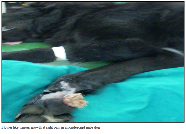
A one and half year old, male non–descript (Bhutia) dog was brought to Institute Referral Veterinary Polyclinics with the history of limping and a unique flower shaped growth in right fore–limb since a week. On physical examination, the growth was found to be pinkish, hairy with irregular contours and tender on palpation, firmly attached to the skin and well demarcated from other tissues (Figure 1).
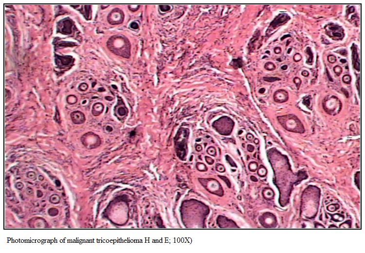
All vital parameters were within the normal physiological range. Thoracic and abdominal survey radiographs revealed no metastatic lesion in lung and other tissues. An excisional biopsy was taken from the mass and submitted for histopathology. Histological examination revealed the presence of multiple islands of hair follicular structures surrounded by the proliferating epithelial cells with hyperchromatic nuclei, scanty eosinophilic cytoplasm. Few cells were having granular cytoplasm and forming cystic spaces. The neoplastic cells were also invading into the surrounding connective tissue, which was showing moderate degree of desmoplastic reaction (Figure 2).Based on clinical picture, macroscopic appearance of lesion, histological characterization and survey radiographic findings, the case was diagnosed as malignant tricoepithelioma. Since surgical excision is reported curative for this type of tumour, it was decided to excise the tumour growth surgically.
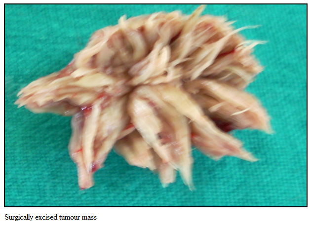
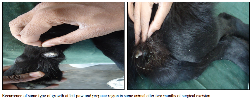
Figure 4-5:Recurrence of same type of growth at left paw and prepuce region in same animal after two months of surgical excision
The animal was prepared for aseptic surgery and premedicated with atropine @ 0.04 mg/kg IM. Anesthesia was then induced with xylazine and ketamine (xylazine @ 1mg/kg and ketamine @ 5mg/kg, both IM). After induction, it was maintained by ketamine and diazepam @ 5mg/kg and 0.5mg/kg body weight respectively, IV. The tumour mass was carefully excised by elliptical incision including 1cm of skin away from the base of the tumour (Figure 3) after achieving surgical plane of anesthesia. The skin was sutured by simple interrupted suture using No.1 polyamide (Linex®) and the excised mass was preserved in Neutral buffer formalin solution and submitted for histopathology. Post– operatively ceftriaxone (20mg/kg, BD, IM) was given for seven days After two months of surgical treatment, the same animal presented with same type of growths in prepuce and left paw region (Figure 4 & 5). Radiography of thorax was performed and revealed no evidence of metastasis. Excisional biopsy of these growths also confirmed the same tumour type and consequently the tumour growths were surgically removed as described earlier using the same anesthetic protocol. Since the tumour was multicentric and malignant in nature, it was decided to combine chemotherapy along with surgical removal. Cyclophosphamide (Cyphos®) @ 50 mg/m2 IV was given at weekly interval for 3 weeks. The skin sutures were removed on 10th postoperative day and the animal showed no recurrence of tumor at different surgical site during the follow–up period of two years (Figure 6).
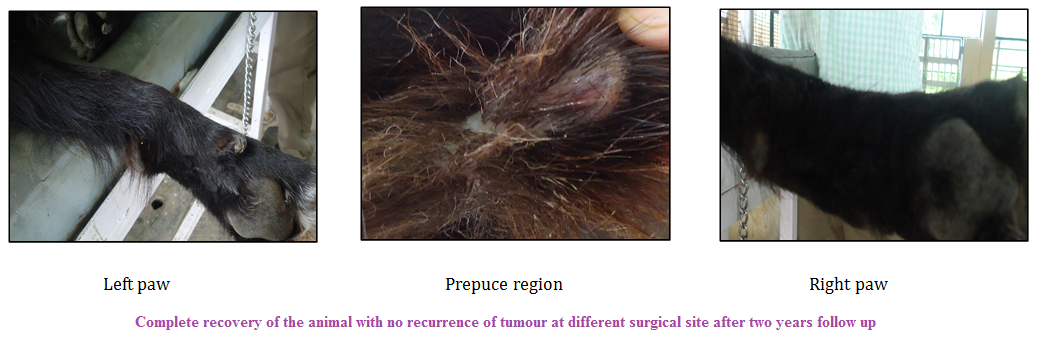
Figure 6:Complete recovery of the animal with no recurrence of tumour at different surgical site after two years follow up
Malignant tricoepithelioma is a malignant tumour with metrical and inner root sheath differentiation, which may metastasize. The tumour is seen as a nodular infiltrative mass involving dermis and subcutaneous tissue, and is indistinguishable grossly from other invasive skin tumours (Scott and Aderson, 1991). However due to its low incidence, the clinical picture has not been well described in literature. Earlier workers also recorded trichoepithelioma in a dog which revealed features like islands of basal cells with keratinized centers of horn cysts and few rudimentary hair follicles (Anjaneyulu and Haragopal, 2002; David and Stephen, 2007).
Basset Hounds and English Springer Spaniels have increased risk for developing multiple tricoepithelioma (Kahn and Scott line, 2005), but not the malignant form. Probably this might be the first report. Animals that develop tumour are prone to develop additional lesions at other sites (Kahn and Scott line, 2005) and its incidence is about 6%. The multicentric occurrence in this case may be due to the same reason. Cyclophosphamide is commonly used in skin tumors such as epitheliotropic lymphoma (Tzannes et al., 2008), subcutaneous haemangiosarcoma (Bulakowski et al., 2008), mast cell tumors (Gerritsen et al., 1998) and mammary gland tumour (Rutteman et al., 2001). Rather using high dose at 21 days interval, we used low dose at week interval. Clinical recovery and absence of tumour recurrence after surgico–chemotherapy indicates that this combination may be useful in preventing tumour recurrence and need further evaluation.
ACKNOWLEDGEMENT
The authors are highly thankful to the Incharge, Institute Referral Polyclinics; Head, Division of Surgery and Director, Indian Veterinary Research Institute, Izatnagar, Uttar–Pradesh, India for providing necessary facilities to carry out this work.
CONFLICTOF INTEREST
The authors declare that they have no conflicts of interests with respect to their authorship or the publication of this article.
REFERENCES
Anjaneyulu Y, Haragopal V (2002). Trichoepithelioma in a dog. Ind. Vet. J. 79: 1198
Bulakowski EJ, Philibert JC, Siegel S, Clifford CA, Risbon R, Zivin K, Cronin KL (2008). Evaluation of outcome associated with subcutaneous and intramuscular hemangiosarcoma treated with adjuvant doxorubicin in dogs: 21 cases (2001 – 2006). J. Am. Vet. Med. Assoc. 233(1): 122 – 128
http://dx.doi.org/10.2460/javma.233.1.122
PMid:18593321
David MV, Stephen JW (2007) Tumours of skin and subcutaneous tissue. In: MacEven EG, Withrow SJ, editors. Small Animal Clinical Oncology, Elsevier Health Sciences, p. 389 – 390.
Gerritsen RJ, Teske E, Kraus JS, Rutteman GR (1998). Multi–agent chemotherapy for mast cell tumours in the dog. Vet. Q. 20(1): 28 – 31.
http://dx.doi.org/10.1080/01652176.1998.9694832
PMid:9477533
Goldschmidt MH, Hendrick MJ (2002). Tumors of the skin and soft tissues. In: Meuten DJ, (ed.). Tumors in domestic animals, 4th ed. pp. 60, Iowa State Press, Iowa, USA.
http://dx.doi.org/10.1002/9780470376928.ch2
Kahn CM, Scott line (2005). The Merk Veterinary Manual. 9th ed. Merch and Co., Inc, NJ, USA
Rutteman GR, MacEwen EG, Withrow SJ (2001). Tumors of the mammary gland. In: MacEwen EG and Withrow SJ, editors. Small Animal Clinical Oncology, 3rd ed. W. B. Saunders: London
PMCid:PMC1505860
Scott DW, Aderson WI (1991). Canine hair follicle neoplasm: A retrospective study of 80 cases (1986–1987). Vet. Dermat. 2: 143–150.
http://dx.doi.org/10.1111/j.1365-3164.1991.tb00125.x
Tzannes S, Ibarrola P, Batchelor DJ, Burrow RD, Blackwood L (2008). Use of recombinant human interferon alpha–2a in the management of a dog with epitheliotropic lymphoma. J. Am. Anim. Hosp. Assoc. 44(5): 276 –282.
PMid:18762565
Wallace BM (2002) Cancer in Dogs and Cats: Medical and Surgical Management. 2nd ed. Teton New Media: Wyoming.


