Advances in Animal and Veterinary Sciences
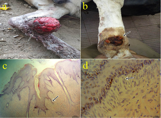
(a-b): Gross picture of the fibroblastic type of sarcoid showing fleshy ulcerated surface (arrow) (1a). Verrucous lesions showing a warty appearance with variable degrees of flaking and scaling of their surfaces (arrow) (1b). (c-d): Photomicrograph of H&E stained equine sarcoid showing epidermal acanthosis, hyperkeratosis, and hyperplasia with long rete pegs (arrow) (bar 200µm) (1c) and picket fence formation (arrow) (bar 20µm) (1d).
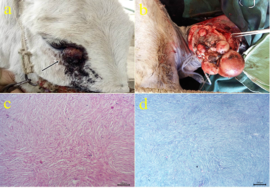
(a-b): Gross picture of fibroma showing nodular raised mass with the alopecic surface at periocular skin (arrow) (2a) and Pedunculated polypoid mass on the skin at the medial aspect of the forearm (1b). (c): Photomicrograph of H&E stained fibroma showing interlacing bundles of spindle-shaped fibroblast cells containing collagen fibers. (bar 100µm) (d): Photomicrograph of Masson’s trichrome-stained collagen of fibroma. (bar 100µm).
(a): Gross picture of SCC in male donkey showing Cauliflower-like mass at periocular and eyelid skin (arrow). (b-c): Photomicrograph of H&E stained SCC showing nests of neoplastic epithelial cells (arrow) (bar 100µm) (3b), obvious mitotic figures (arrow) surrounded by lymphocytes (arrowhead) (bar 20µm) (3c). (d) Positive cytoplasmic expression of CK5 within the invaded epithelium (arrowhead) of IHC counterstaining with Mayer’s hematoxylin (bar 20µm)
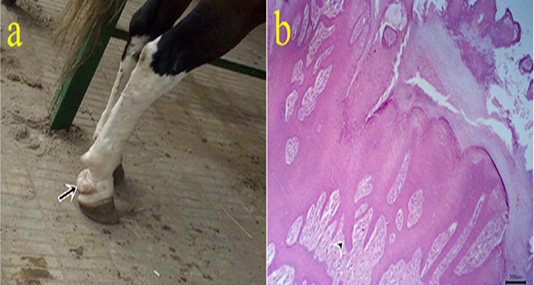
(a): Gross picture of papilloma in male horse showing white firm mass at the caudal aspect of pastern joint (arrow). b: Photomicrograph of H&E stained papilloma showing hyperkeratosis and acanthosis with finger-like papillae (arrowhead). (bar 200µm)
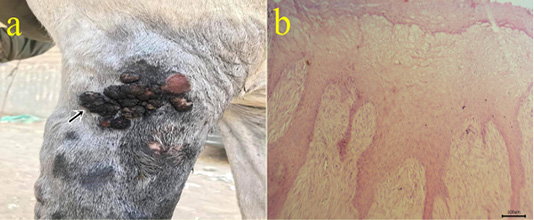
(a): Gross picture of fibropapilloma showing wart-like masses at the lateral aspect of arm region (arrow). (b): Photomicrograph of H&E stained fibropapilloma showing hyperkeratosis , acanthosis with hypercellularity of dermal fibroblasts in the form of whirls (bar 100µm).
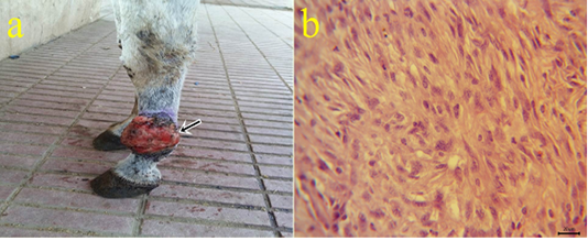
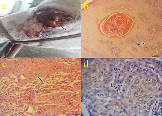
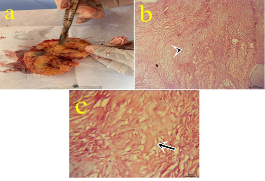
a: Gross picture of fibrosarcoma showing grayish red elevated mass with an ulcerated surface at the skin of the fetlock region (arrow). b: Photomicrograph of H&E stained fibrosarcoma showing pleomorphic fibroblasts with whirls (bar 20µm).
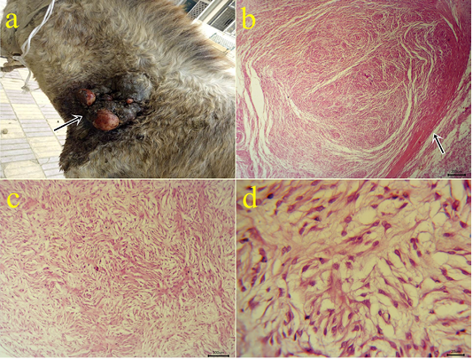
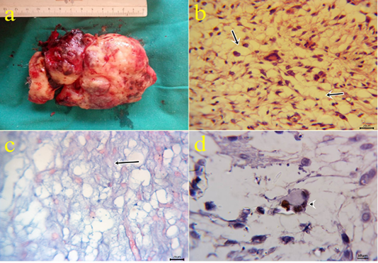
a: Gross picture of myxoma showing a solid neoplastic mass on the skin of the neck in the male donkey. Fig.(7b-7d): Photomicrograph of H&E stained myxoma showing capsulated neoplasm (arrow) (7b), different shapes of embryonic fibroblasts with processes and faint basophilic secretions (7c-7d). (bar 200,100,20 µm) respectively.
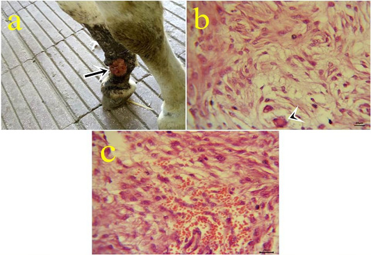
a: Gross picture of myxosarcoma showing solid, ulcerated grayish neoplastic mass at the fetlock joint in donkey (arrow). Fig.(8b-8c): Photomicrograph of H&E stained myxosarcoma showing pleomorphic cells and giant cells (arrowhead) (8b) (bar 20µm). Hypercellularity of embryonic fibroblasts with hemorrhage (8c) (bar 20µm).
a: Gross picture of myxoid liposarcoma showing large glistening slimy neoplastic excised mass from forelimb. Fig.(9b): Photomicrograph of H&E stained myxoid liposarcoma showing poorly differentiated fat cells (arrows) embedded in mucoid substance with undetectable collagen contents and variable sizes giant cells (bar 20µm). Fig.(9c): positive bluish myxoid background (arrow) using alcian blue staining (bar 20µm). Fig.(9d): MDM2 positive nuclear expression within the giant cells (arrowhead) of IHC counterstaining with Mayer’s hematoxylin (9d). (bar 10µm)
a: Gross picture of teratoma showing enlarged firm yellowish excised ovarian mass from 7 years old mare. Fig.(10b-10c): Photomicrograph of H&E stained teratoma showing chondrocytes (arrowhead) (10b) (bar 100µm) and bone spicules (arrow) (10c). (bar 20µm)


