Advances in Animal and Veterinary Sciences
Research Article

Morphology of the Air Sacs in Crimson Rosella (Platycercus elegans) Parrots
Pamela Bejdić1*, Nejra Hadžimusić2, Sabina Šerić-Haračić3, Alan Maksimović4, Ismar Lutvikadić4, Amina Hrković-Porobija5
1Department of Anatomy and histology with embryology, Veterinary Faculty, University of Sarajevo, Zmaja od Bosne 90, 71000 Sarajevo, Bosnia and Herzegovina; 2Department of Pathophysiology, Veterinary Faculty, University of Sarajevo, Zmaja od Bosne 90, 71000 Sarajevo, Bosnia and Herzegovina; 3Department of Animal Health Economics, Veterinary Faculty, University of Sarajevo, Zmaja od Bosne 90, 71000 Sarajevo, Bosnia and Herzegovina; 4Department of Surgery, Oncology and Ophthalmology, Veterinary faculty, University of Sarajevo, Zmaja od Bosne 90, 71000 Sarajevo, Bosnia and Herzegovina; 5Department of Chemistry, Biochemistry and Physiology, Veterinary Faculty, University of Sarajevo, Zmaja od Bosne 90, 71000 Sarajevo, Bosnia and Herzegovina.
Abstract | The research was conducted with the aim to investigate the morphology of air sacs system in Crimson Rosella (Platycercus elegans) parrots. Five adult birds, two males and three females were used in this study. The lungs and air sac system were injected via trachea with 26% solution of Vinylite mass. The obtained cast showed that these parrots have nine air sacs. The clavicular air sac was the only unpaired, while the cervical, cranial thoracic, caudal thoracic and abdominal air sacs were paired. The morphology of the air sacs was generally similar to that reported in other bird species, however, some specific features were identified. As most prominent among them were a partial fusion of the cervical air sacs, communication between the left and right subpectoral and perirenal diverticula and connection between the claviclar and cranial thoracic air sacs. The present investigation provided detailed and comprehensive data about the morphology of air sacs system in these parrot species and these findings will be very useful for future clinical examination and treatment of this birds.
Keywords | Anatomy, Cast corrosion technique, Air sacs, Diverticula
Received | July 22, 2021; Accepted | August 02, 2021; Published | September 25, 2021
*Correspondence | Pamela Bejdić, Department of Anatomy and histology with embryology, Veterinary Faculty, University of Sarajevo, Zmaja od Bosne 90, 71000 Sarajevo, Bosnia and Herzegovina; Email: pamela.bejdic@vfs.unsa.ba
Citation | Bejdic P, Hadzimusic N, Seric-Haracic S, Maksimovic A, Lutvikadic I, Hrkovic-Porobija A (2021). Morphology of the air sacs in crimson rosella (platycercus elegans) parrots. Adv. Anim. Vet. Sci. 9(11): 1959-1963.
DOI | http://dx.doi.org/10.17582/journal.aavs/2021/9.11.1959.1963
ISSN (Online) | 2307-8316; ISSN (Print) | 2309-3331
Copyright © 2021 Bejdic et al. This is an open access article distributed under the Creative Commons Attribution License, which permits unrestricted use, distribution, and reproduction in any medium, provided the original work is properly cited.
Introduction
The Crimson Rosella (Platycercus elegans) is a medium-sized, colourful parrot native to eastern and south-eastern Australia (Penck et al., 1995). Today there is a rising trend of keeping these parrots in captivity as pets. As a consequence of that there have been reports from different veterinary clinics regarding the treatment of respiratory system diseases in these birds (Palmieri et al., 2011; Talbot et al., 2018). However, the literature data about the anatomy of the respiratory system in these parrots still lacking. Different research confirms that the avian respiratory system is structurally and functionally one of the most complex among the vertebrates (Maina, 2008; 2015). In birds, this system consists of a gas exchange part that represents the lungs and parabronchi and a ventilation part that represent the air sacs. The morphology of the air sacs system has been described in many domestic and wild bird species (Getty 1975; Nickel et al., 1977; King and McLelland, 1984; Hamlet and Fisher, 1967; Kloek and Casler 1972; Dyce et al., 2002; Demirkan et al., 2006; Onuk et al., 2009; Orhan et al., 2009, Sawad and Udah 2012; El-Bably et al., 2014; Pandey et al., 2015; Ragab and Reem, 2016; El-Sayed and Hassan, 2019; Bejdić et al., 2021). These research indicate that the number, size and shape of the air sacs and their diverticula are quite different among bird’s species. Depending on the species, it has been found that birds have from six to eleven air sacs (Getty 1975; Nickel et al., 1977; Kadosaki 1977;1978; King and McLelland 1984; Baumel et al., 1993, Sawad and Udah 2012; Ragab and Reem, 2016; König et al., 2016). Considering the above mentioned, the aim of this study was to morphologically describe the air sacs system in Crimson Rosella parrots and provide useful data for the veterinary practitioners and researchers.
Material and methods
Five adult Crimson Rosella (Platycercus elegans) parrots were used in this study, two males and three females. All birds were collected after they have died at the clinic of the Veterinary faculty, University of Sarajevo during the last four years. None of the birds had respiratory system diseases. Two females died from egg binding, one male had heart failure, and one male and one female had a traumatic, inoperable injury. All birds were transferred to the Department of Anatomy and histology with embryology, where a detailed morphological examination of the air sacs has to be performed. For the purpose of this study, we applied the corrosion cast technique according to Tompsett DH. 1970. As injection mass, we used the 26% solution of Vinylite dissolved in acetone p.a. The prepared mass was additionally coloured in yellow (BIODUR® Colour Paste Corn Yellow AC 53). The Vinylite mass was injected into the trachea by plastic syringe with needle. During the injection, the pressure on the abdominal wall was constantly monitored by manual palpation. When the desired pressure was achieved, the injection was stopped and the trachea was enclosed by surgical forceps. The birds were emerged into the tap water for 24 hours and later placed into the device for soft tissue maceration of biological specimens (Schaerer, Weber-Bern). After 21 days, the specimens were washed in tap water and left to dry for 24 hours. At the end of this process, we got the full cast of the bronchi and air sacs with the preserved skeleton. For better visualization of the casted structures, some of the bones were removed. These specimens are used for precise determination of the position, shape and size of the air sacs and their diverticula. For proper Latin terminology, we used the Nomina Anatomica Avium (Baumel et al., 1993).
Results and Discussion
The obtained Vinylite casts show that Crimson Rosella parrots (Platycercus elegans) have nine air sacs. The clavicular air sac (saccus clavicularis) was the only unpaired, while the cervical (saccus cervicalis), cranial thoracic (saccus thoracicus cranialis), caudal thoracic (saccus thoracicus caudalis) and abdominal air sacs (saccus abdominalis) were paired (Figure 1). There were no differences between male and female birds.
The clavicular air sac (saccus clavicularis) lies in the cranial part of the thoracic cavity, between the heart, pectoral girdle and sternum. The six diverticula, two intra-thoracic and four extra-thoracic separated from this sac. A similar position of the clavicular air sac and its diverticula has been reported in domestic and some wild bird species (Getty 1975; Nickel et al., 1977, King and McLelland 1984, Baumel et al., 1993, Dyce et al., 2002, König et al., 2016).
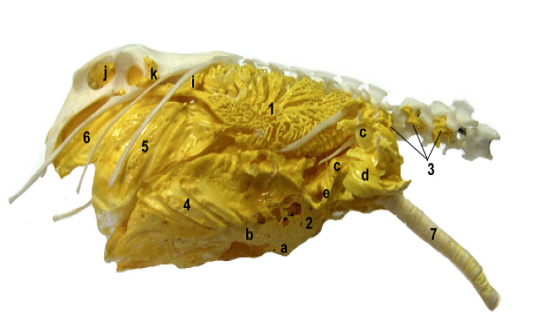
Figure 1: A lateral view on in situ casted the respiratory system in Crimson Rosella parrots showing: 1. bronchi, 2. clavicular air sac with it’s: a) diverticula cardiaca, b) diverticula sternalia, c) diverticula subpectorale, d) diverticula suprahumerale, e) diverticula axillare and f) divertiuclua subscapulare, 3. cervical air sac with its diverticula vertebrale, 4. cranial thoracic air sac, 5. caudal thoracic air sac, 6. abdominal air sac with its cranial (i) and caudal (j) diverticula perirenalia and diverticula femoralia (k) and trachea (7).
The intra-thoracic diverticula were diverticula cardiaca and sternalia, and the extra-thoracic were diverticulum subpectorale, suprahumerale, axillare and subscapulare (Figure 1). The diverticula cardiaca emerge from the caudal side of the clavicular air sac and covers the base of the heart and atrial wall. The impressions of the large blood vessels were visible on the casts. The diverticula sternalia separtes from the caudoventral and lateral side of the clavicular air sac and aerates the sternum and sternal part of the ribs. Along with the level where sternum and the ribs joints, diverticula sternalia connects with the cranial thoracic air sac (Figure 1 and 2). This connection between the clavicular and cranial thoracic air sacs so far has been recorded in a Rosy-faced parrots (Bejdić et al., 2021), Hooded crow (El‐Sayed and Hassan, 2019), Common grackle, Cardinals, Rufous-sided Towbees, Song Sparrow and some individual Old World songbirds (Kloek and Casler, 1972), but never in domestic birds (King and McLelland 1984, Baumel et al., 1993, König et al., 2016). The position and shape of this connection was very similar to that recorded in Rosy-faced parrots (Bejdić et al., 2021), what indicates that this can be common characteristics of the air sac system in parrot species. From opposite, craniolateral side of the clavicular air sac emerge a large, triangular subpectoral diverticula (Figure 1 and 2). Above the trachea, the left and right diverticula subpectoralia connects through one wide, semicircular, bridge-like extension (Figure 3). This connection has not been reported previously in other bird species. Regarding the other characteristics like as the position, shape and size of these diverticula, they were in accordance with findings in other bird species (Hamlet and Fisher, 1967, Kloek and Calser 1972, Dyce et al., 2002; Orhan et al., 2009, Onuk et al., 2009, El-Bably et al., 2014, Ragab and Reem, 2016, El-Sayed and Hassan 2019, Bejdić et al., 2021). Laterally for clavicular air sac separates the suprahumeral diverticula. These diverticula have a large curved shape and goes around the proximal extremity of the humerus (Figure 1 and 2). The small axillary diverticula emerge caudoventrally from them and these aerate the coracoid bone (Figure 1). Above axillar separates the subscapular diverticula who gives many small extensions that occupy the spaces between the first two thoracic vertebra and vertebral part of first two ribs. In domestic fowl and some wild bird species these diverticula were described as simple, leaf-like structures who lies between the scapula and cervical air sacs (King and McLelland, 1984; Demirkan et al., 2006; Orhan et al., 2009; El-Sayed and Hassan 2019). The present research showed that in Crimson Rosella parrots these diverticula were structurally much more complex and they were similar to those described in Rosy-faced Parrots (Bejdić et al., 2021).
The cervical air sacs (saccus cervicalis) were the smallest of all and situated above the trachea, where extending from the second thoracic to last three to four cervical vertebrae (Figure 1). The medial wall of these sacs partially fused so they had a shape of the “X” letter (Figure 3). From the caudodorsal side of the cervical air sacs emerge small diverticula vertebralia who occupy the spaces between first two thoracic to last four cervical vertebrae (Figure 1). The terminal parts of these diverticula enters into the vertebral and transversal canal and establish the connection between the left and right side. In many bird species, the cervical air sacs were described as a paired with diverticula verterbralia and intermuscularia (Kloek and Casler 1972; Kadosaki 1977; 1978; King and McLelland, 1984; Baumel et al., 1993; Dyce et al., 2002; Maina 2008; Onuk et al., 2009; El-Bably et al., 2014; König et al., 2016; Bejdić et al., 2021). In the present investigation, these sacs were also paired, but partially fused and without the diverticula intermuscularia. The complete fusion of these sacs was recorded in Long-Legged Buzzard and Japanese Quail (Orhan et al., 2009; Sawad and Udah, 2012), while in Turkey (Ragab and Reem, 2016) and Goose (Onuk et al., 2019) the unpaired cervical air sac additionally fused with clavicular air sac forming the one cervicoclavicular sac.
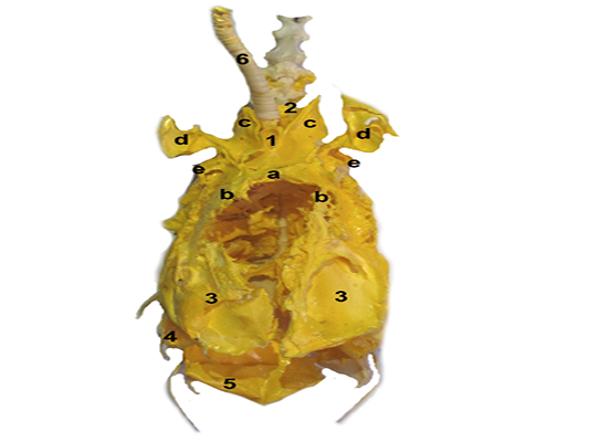
Figure 2: A ventral view on corroded in situ specimen with visible: 1. clavicular air sac, a) diverticula cardiac, b) sternalia, c) subpectorale, d) suprahumerale, e) axillare, 2. cervical air sac with its diverticulum vertebrale, 3. cranial thoracici air sac, 4. caudal thoracic air sac, 5. abdominal air sac with its cranial (i) and caudla (j) diverticula perirenalia, femoralia and 6. trachea

Figure 3: A cranial view on partially fused (*) the cervical air sacs (1), diverticula vertebralia (a), 2. trachea, 3. clavicular air sac, subpectoral (c) and suprahumeral (d) diverticula.
The cranial thoracic air sacs (saccus thoracicus cranialis) were oval and larger than the caudal thoracic air sacs. Their dorsal margin lies at the level where vertebral and sternal part of the ribs joins (Figure 1). Ventrally these sacs connects with the clavicular air sac through theirs sternal diverticula of and together they aerate the sternum and ribs as we described it above (Figure 1 and 2). In many bird species, the cranial thoracic air sacs were described as smaller than the caudal and both do not give any diverticula or aerate the bones what is opposite to our findings (Getty, 1975; Nickel et al., 1977; King and McLelland 1984; König et al., 2016). At other side, the similar observations have been recorded in Rosy-faced parrtos (Bejdić et al., 2021), Long-legged Buzzard (Orhan et al., 2009), Mallard (Dermikan et al., 2006) and Golden Peking duck (El-Bably et al., 2014). Additionally, the diverticulum subcardiale and supracardiale have been described in Long-legged Buzzard, but we did not find these in the present study (Orhan et al., 2009).

Figure 4: A ventral view on abdominal air sac (1) and diverticula perirenalia cranialis (a), caudalis (b) and femoralia (c).
The caudal thoracic air sacs (saccus thoracicus caudalis) were located between the cranial thoracic and abdominal air sac. Their lateral surface lies on vertebral parts of the last three ribs (Figure 1). These sacs do not have any diverticula or aerate the bones, which is in accordance with previous findings in domestic and other bird species (Kloek and Casler 1972; Kadosaki 1977; 1978; Baumel et al., 1993; Dermikan et al., 2006; Onuk et al., 2009; Orhan et al.,. 2009; El-Bably et al., 2014; Ragab and Reem, 2016; König et al., 2016).
The abdominal air sacs (saccus abdominalis) were the largest and they occupied the dorsolateral side of coelomic cavity, extending from the caudal margin of the lungs to the cloaca (Figure 1 and 4). The right abdominal sac was slightly larger than the left. The diverticula perirenalia and femoralia separate from these sacs. On the casts it can be distinguished the cranial and caudal diverticula perirenalia (Figure 4). The cranial diverticula perirenalia emerge from the dorsal side of the abdominal sac at the level of last thoracic and first lumbar vertebra. Medially, the left and right diverticula join and aerate the cranial part of the synsacrum (Figure 4). The caudal diverticula perirenalia separates also from the dorsal side of the abdominal air sac, but at the level of the hip joint. These diverticula were wide, long and lie above the kidney where aerate the caudal part of synsacrum. The femoral diverticula emerge from the ventrolateral side of abdominal air sacs in a front of the hip joint (Figure 1 and 4). They were small, semicircular and follow the margin of the acetabulum. In previous reports the perirenal diverticula were generally described as several extensions who emerge dorsally from these sacs and go between the kidney and synsacrum where invade the adjacent vertebrae and pelvic girdle (Getty, 1975; Nickel et al., 1977; King and McLelland, 1984; König et al., 2016; El‐Sayed and Hassan, 2019), but there is no detailed description of the same. Also, in literature, we did not find data about the presence of the connection between the left and right diverticula. This research provides a detailed description of all air sacs, their diverticula, and their interconnections.
CONCLUSION
The morphology of the air sacs system in Crimson Rosella parrots was very similar to that reported in other bird species, however, some specific features were identified. As most prominent among them were a partial fusion of the cervical air sacs, communication between the left and right subpectoral and perirenal diverticula and connection between the claviclar and cranial thoracic air sacs. The present investigation provided detailed and comprehensive data about the morphology of air sacs system in these parrot species and these findings will be very useful for future clinical examination and treatment of this birds.
conflict of interest
Authors declare that there is no conflict of interest.
Acknowledgement
The authors are grateful to Jasmina Mulaosmanović for the professional English review and editing.
authors contribution
All authors have equally contributed to this manuscript.
References


