Advances in Animal and Veterinary Sciences
Research Article
Advances in Animal and Veterinary Sciences 1 (3S): 17 – 20Special Issue–3 (Epidemiology and Animal Disease Investigations)
Natural Outbreak of Newcastle Disease in Turkeys and Japanese Quails Housed Along With Chicken in a Multi–Species Poultry Farm in Northern India
Vasudevan Gowthaman, Shambu Dhayal Singh*, Rajamani Barathidasan, Anjaneya Ayanur, Kuldeep Dhama
-
Avian Diseases Section, Division of Pathology, Indian Veterinary Research Institute, Izatnagar, Bareilly–243122, Uttar Pradesh, India
*Corresponding author:sdsingh2005@rediffmail.com
ARTICLE CITATION:
Gowthaman.V, Singh SD, Barathidasan R, Ayanur A and Dhama K (2013). Natural outbreak of Newcastle disease in turkeys and japanese quails housed along with chicken in a multi–species poultry farm in northern India. Adv. Anim. Vet. Sci. 1 (3S): 17 – 20.
Received: 2013–11–18, Revised: 2013–12–29, Accepted: 2013–12–30
The electronic version of this article is the complete one and can be found online at
(
http://nexusacademicpublishers.com/table_contents_detail/4/192/html
)
which permits unrestricted use, distribution, and reproduction in any medium, provided the original work is properly cited
ABSTRACT
Newcastle Disease (ND) is a highly infectious and contagious disease of domestic poultry and wild birds that could cause high morbidity and mortality. Newcastle Disease virus (NDV) infects wide variety of hosts including turkeys and quails. ND is highly endemic in Indian poultry industry. Frequent episodes of disease occur in domestic birds and it is capable to infect other bird species which kept alongside with poultry. There were no recoded reports on the occurrence of NDV in the commercial chickens farmed along with multi species, especially, turkeys and Japanese quails. A disease investigation was undertaken in a multi species poultry farm with the history of morbidity >30% and mortality (>20%) with unusual clinical manifestations of the central nervous system dysfunction in turkeys and Japanese quails kept along with poultry. Systematic necropsy examination was carried out in dead turkeys and Japanese quails and visceral organs were collected for virus isolation and histopathology. Cloacal swabs were also collected from apparently healthy chicken flocks. Virus isolation, Mean Death Time (MDT) assay and histopathology were carried out as per standard procedures. All infected turkeys and Japanese quails showed general weakness, loss of appetite, decrease in egg production and central nervous system (CNS) signs like torticollis, uncoordinated gait and backward movements, abnormal positioning of the head and neck such as opisthotonus. Postmortem examination and histopathology revealed prominent vascular and CNS alterations. ND virus was isolated from all the pooled samples from turkeys and Japanese quails and 3 cloacal swabs from chickens. The MDT of the all isolates from turkeys and quails found to be 38– 60 hours, whereas for chicken isolates the MDT exceeded 90 hours. This confirms the velogenic nature of the isolated ND virus in turkeys and Japanese quails.
INTRODUCTION
Newcastle Disease (ND) is a highly infectious and contagious disease of domestic poultry and wild birds that could cause high morbidity, mortality and severe economic losses to the poultry industry worldwide (Sonaiya and Swan 2005; Leuck et al., 2004). It is of great concern to other domestic species maintained along with commercial poultry. ND is caused by avian paramyxoviruses belongs to the genus Avulavius and family Paramyxoviridae which included in the order Mononegavirales (Lamb et al., 2005). There are eleven serotypes of avian paramyxoviruses (APMV) designated APMV–1 to APMV–11 (Tiana et al., 2012) have been reported so for. Among which APMV–1 infects wide variety of hosts including turkeys and quails. NDV is a single stranded, non segmented, negative–sense RNA viruse with helical capsid symmetry (Wise et al., 2004; Rue et al., 2011; Samuel et al., 2013). Kaleta and Baldauf (1988) reported that at least 241 species of birds from 27 of the 50 orders of birds are susceptible to ND virus (NDV). It is one of the OIE listed disease having trade importance (OIE, 2005, 2008). Classically, the virulence of NDV is assessed by intracerebral pathogenicity index (ICPI) and/or intravenous pathogenicity index (IVPI), and/or mean death time (MDT) in embryonating eggs. The virus has been classified into 5 strains based on its pathogenicity viz., 1) Viscerotropic velogenic 2) Neurotrophic Velogenic 3) Mesogenic 4) Lentogenic 5) Asymptomatic enteric NDV (Beard and Hanson, 1984). The virus was first reported In India by Edwards in the year 1928 (Edwards, 1928) in Ranikhet region and it is popularly called as Ranikhet disease (RD) in India. ND is highly endemic in Indian poultry industry despite the widespread use of vaccines (Gowda, and Eswaran, 1992; Sulochana and Mathew, 1991). Frequent episodes of disease occur in domestic birds and it is capable to infect other bird species which kept alongside with chickens (Ucan et al., 2002). Newcastle disease has been reported frequently in turkeys (Graham et al., 1996; Alexander et al., 1998) and sporadically in Japanese quails worldwide (Czirják et al., 2007). The Japanese quails are usually acting as carriers of NDV and resistant to NDV compared to chickens. There were no recoded reports on the occurrence of NDV in the commercial multi–species poultry farms. In the recent years, turkey and quail industry has been flourishing in a large commercial scale. Therefore, it is necessary to study the epidemiology and pathology of NDV in other species of poultry housed along with chickens. This paper describes an outbreak of ND in turkeys and Japanese quails (Coturnix coturnix japonica), with confirmation of the aetiology and pathology in a multi–aged and multi–species poultry farm which routinely practices NDV vaccination.
MATERIALS AND METHODS
History
The clinical history includes morbidity (>30%) and mortality (>20%) with signs of central nervous system dysfunction observed in Japanese quails and turkeys in a multi–species poultry breeding farm, which housed about 1.5 lac Japanese quails and 50,000 breeding turkeys and chickens. Antibiotic treatment for four consecutive days failed to check the mortality. The age of the affected quail flock was 22 weeks, turkeys were 18 weeks and chickens were 35 weeks–old. The commercial chickens remained healthy despite of severe disease outbreak in turkeys and quails. The farm was located in Mewat district of Haryana state in Northern India. The birds were vaccinated against egg drop syndrome–76 (EDS–76), Infectious Bronchitis (IB) and Marek’s disease (MD) at the earlier age, and recently, 46 days prior to the outbreak all the birds were vaccinated against Newcastle disease (NDV–laSota). An investigation was undertaken to find out the cause of increased mortality in the affected farm.
Necropsy and Sampling
Systematic necropsy examination was carried out in dead turkeys and Japanese quails; trachea, lung, spleen, intestines, proventriculus, and brain tissues were collected aseptically from the dead and euthanized birds for virus isolation as well as in 10% buffered formal saline for histopathology. Thirty fecal samples (10 each from turkey, quails and chickens) were collected for parasitological examination. The cloacal swabs were randomly collected from chickens for virus isolation.
Virology
One hundred milligram of pooled brain, trachea and spleen tissues samples from twenty birds (10 each from turkey and quails) were homogenized in 1 ml 1X PBS in a sterile pestle and mortar to make a 10% suspension. The suspension was transferred to a centrifuge tube and clarified at 12,000 rpm for 10 minutes to remove extraneous materials and the isolation was confirmed by HA test. To the clear tissue homogenate 1/10 volume of 1X antibiotic mixture was added and incubated at 37oC for one hour finally the supernatants were filtered through a 0.45µm filter and filtrates were inoculated into 10 day–old embryonated chicken eggs via allantoic route for Newcastle disease virus (NDV) isolation as described earlier (OIE, 2008). The cloacal swabs were also processed as per the standard methods (Alexander, 1998). The HA–positive samples were assayed by hemagglutination inhibition (HI) test with NDV–specific antiserum.
Mean Death Time Assay
MDT assay was carried out as per the standard procedure described in elsewhere (Alexander, 1998).
Histopathology
The tissue samples were collected in 10% buffered formal saline fixed for 48–72 hours. After fixation, the tissue samples were processed further for routine paraffin embedding of tissue. Briefly, the samples of desired thickness were washed in running tap water overnight. The tissues were dehydrated in increasing concentrations of alcohol ranging from 70%, 80%, 90% and absolute alcohol, acetone, cleared in xylene and embedded in paraffin wax. The tissues were sectioned to 4–5 micron thickness and sections were taken on poly–L lysine precoated slides. After drying, the sections were stained with haematoxylin and eosin with slight modification as per the standard procedures (Luna, 1968).
RESULTS
Clinical Signs
All infected turkeys and Japanese quails showed general weakness, loss of appetite, decrease in egg production and CNS signs like torticollis, uncoordinated gait and backward movements, abnormal positioning of the head and neck such as opisthotonus. In turkeys, nervous signs, such as head twitches and tremor, backward movements and paralytic syndrome were severe when compared to quails; majority of the quails showed torticollis and uncoordinated movements only. In addition, turkeys had conjunctivitis, severe periocular edema and severe respiratory distress. No such signs were observed in Japanese quails except mild respiratory distress. Besides, majority of the birds showed haemorrhagic or greenish diarrhoea. The chickens housed in the adjacent sheds remained apparently healthy.
Postmortem Lesions
Post mortem examination revealed marked to severe and widespread congestion of internal organs in Japanese quails, where as in turkeys no visceral congestion was evidentexcept congestion of the meninges. Diffuse catarrhal tracheitis, edematous and haemorrhagic lungs were the prominent lesions observed in turkeys, but no significant respiratory lesions were found in Japanese quails. The air sacs were cloudy in turkeys but remained clear in Japanese quails. The spleens showed marked atrophy and mottling in both the species. The intestine of turkeys showed catarrhal enteritis, whereas in Japanese quails intestine showed severe and diffuse heamorrhages/haemorrhagic ulcers in all the birds examined. Haemorrhages at the tip of the proventricular glands found in few quails.
Histopathology
The most significant histopathological changes revealed severe CNS involvement in both the species. The brain changes consist of a nonsuppurative encephalomyelitis, gliosis, perineuronal edema, neuronal shrinkage and necrosis. All the changes e were present in both the species with varying severity. The liver and kidney of Japanese quails showed vascular changes of congestion and haemorrhages; multifocal necrosis; whereas in turkeys the lesions were less severe. Marked to sever lymphocytic infiltrations, haemorrhage and edema was observed in bronchiolar area as well as air and blood capillaries of the lungs of turkeys. These changes were mild in Japanese quails. Lympocytolysis and lymphoid atrophy/ depletion were the major findings observed in the spleen of both Japanese quails and turkeys. The intestines showed sloughing of the mucosa, vascular engorgement in the tips of the villi, marked to severe haemorrhages and infiltration of mononuclear cells in the lamina propria in both the species; the haemorrhages were prominent in Japanese quails. The pancreas of turkeys showed lymphocytic aggregates.
Bacteriology and Parasitological Examination
On bacteriology and parasitological examination, samples were found negative for possible bacterial pathogens/ parasites.
Virus Isolation
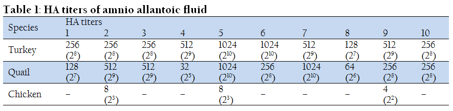
ND virus was isolated form all the pooled samples from turkeys and Japanese quails in the first passage itself. In chickens, virus could be recovered only from 3 samples even after consecutive passages. The mean HA titer was varied between 4(22) to 8(23) in chickens, whereas it remained high in turkeys and quails and varied between 32(25) to 1024 (210). Further confirmation of the APMV –1 was done by neutralization test using ND virus specific antiserum. The details of virus isolation was shown in the Table 1.
MDT
The MDT of the all isolates from turkeys and quails found to be 38– 60 hours, whereas for chicken isolates the MDT exceeded 90 hours. This confirms the velogenic nature of the isolated ND virus in turkeys and Japanese quails. The details of MDT shown in the Table 2.
DISCUSSION
Newcastle disease virus remains as a very significant pathogen for domestic chickens. The virus is capable of infecting other domestic species that are housed along with poultry, but relatively little is known about the dynamics and pathology of the clinical disease in the other species of the birds housed along with poultry. Findings of this study demonstrate that commercial turkeys and Japanese quails exhibit different type of clinical manifestations though the intensity of the disease is severe in both the species. The general clinical signs mainly observed like decrease in egg production, CNS signs like torticollis, greenish diarrhoea in the present study are very typical to that of velogenic ND. Similar findings were reported by previous workers (Wakamatsu et al., 2006; Piacenti et al., 2006; Czirják et al., 2007, Susta et al., 2011; Diel et al., 2012; Brown et al., 2013) in turkeys and quails. Lymphoid depletion and necrosis in lymphoid tissues were observed equally in both the cases. But in our study, there is no respiratory signs were observed in Japanese quails when compared to that of turkeys, which showed severe respiratory distress and histopathological alterations in the respiratory system. These findings correlate with the studies of Momayez et al. (2007), who observed no respiratory signs during the clinical course of NDV in Japanese quails. In the present study, Japanese quails showed severe histopathological alterations in brain and intestines compared to mild patho–morphological alterations in the respiratory system. This suggests that involvement of respiratory system is less pronounced than that of gastro–intestinal and nervous system in Japanese quails. The virus recovered from all the samples of turkeys and quails and the isolates were confirmed as NDV (APMV–1) based on the HA and neutralization tests. The MDT value of all the turkey and quail isolates were less than 60 hrs, confirming them as velogenic NDVs (OIE, 2008), whereas the virus was not recovered in all the cloacal swabs of the healthy chickens and the MDT value of the chicken isolates were greater than 90 hours. This attributed that the recovered chicken isolates could belong to lentogenic and the isolates might belong to the vaccine strain of LaSota virus. Incidence of NDV in Japanese quails is occasional, when compared to incidence of NDV in chickens and turkeys. Japanese quails remain healthy and act as carrier to chicken throughout their life (Lima et al., 2004). Wakamatsu et al., 2006 reported that turkeys are less susceptible to NDV compared to that of chickens. But in the present study, chickens raised in the same farm remained healthy. This could be due to presence of protective antibody titers against NDV in the farm and turkeys and Japanese quails might succumbed to the disease due to vaccination failure or newly evolved strain of NDV or presence of immunosuppressive factors. Ucan et al. (2002) reported that housing quails and chickens together was the possible cause of Newcastle disease spread to chickens. Islam et al. (1994) showed that the enhanced pathogenicity of the mesogenic NDV isolate from quail. In the present study, breeding turkeys housed together with Japanese quails infected with NDV. The critical level of NDV antibodies in turkey flock could not sufficiently protect them against the disease. So breeding turkeys and Japanese quails together might be the source of the disease in turkeys, but further molecular epidemiological studies are needed to confirm the source of transmission. In conclusion the present study demonstrates natural outbreak of Newcastle disease in turkeys and Japanese quails in a multi–species poultry farm. The result also indicates that turkeys and Japanese quails housed in the same farm exhibit different type of clinical disease manifestation to ND. This virus might be emerging in poultry flocks of the country. Further nation–wide epidemiological studies are suggested to know the magnitude of this important poultry virus along with isolation and characterization of field viruses. These studies will clearly help defining the need, if any, for the inclusion of NDV vaccination programmes in turkeys and Japanese Quails.
CONFLICT OF INTEREST
It is to specifically state that "No Competing interests are at stake and there is No Conflict of Interest” with other people or organizations that could inappropriately influence or bias the content of the paper.
REFERENCES
Alexander DJ (1998). Newcastle disease and other avian paramyxoviruses. In: A Laboratory Manual for the Isolation and Identification of Avian Pathogens4th edition. Edited by D.E. Swayne, J.R. Glisson, M.W. Jackwood, J.E. Pearson and W.M. Reed. American Association of Avian Pathologists: Kennet Square. pp 156–163.
Alexander DJ, Morris HT, Pollitt WJ, Sharpe CE, Eckford RL, Sainsbury RMQ, Mansley LM., Gough RE and Parsons G (1998). Newcastle disease outbreaks in domestic fowl and turkeys in Great Britain during 1997. Vet. Rec. 143: 209–212.
http://dx.doi.org/10.1136/vr.143.8.209
PMid:9770762
Beard CW and Hanson RP (1984). Newcastle disease. In M.S. Hofstad, H.J. Barnes, B.W. Calnek, W.M. Reid, H.W. Yoder (eds.). Diseases of Poultry 8th Ed., pp.452–470. Iowa State Univ., Press, Ames.
Brown CC, Sullivan L, Dufour–Zavala L, Kulkarni A, Williams S, Susta L, Zhang J, Sellers H (2013). Comparing presence of avian paramyxovirus–1 through immunohistochemistry in tracheas ofexperimentally and naturally infected chickens. Avian Dis. 57:36–40.
http://dx.doi.org/10.1637/10299-070312-Reg.1
PMid:23678727
Czirják GÁ, Köbölkuti D, Cadar A, Ungvári M, Niculae P (2007). An outbreak of the Newcastle disease in Japanese quail (coturnix coturnix japonica). bulletin usamv–cn, 64/2007 (1–2).
Diel DG, Susta L, Cardenas Garcia S, Killian ML, Brown CC, Miller PJ, Afonso CL. (2012). Complete genome and clinicopathological characterization of a virulent Newcastle disease virus isolate from South America. J. Clin. Microbiol. 50:378–87
http://dx.doi.org/10.1128/JCM.06018-11
PMid:22135263 PMCid:PMC3264182
Edwards, J.T. (1928). Annual Report Imp. Inst. Vet. Res. Mukteswar, India 1928, pp.14–15.
Gowda SRN and Eswaran R (1992). Ranikhet disease in pigeons: Clinicopathological studies. Ind. J. Vet. Pathol., 16 : 95–97.
Graham DA, Connor TJ, McCullough SJ, McKillop ER, Alexander DJ, Manvell RJ, Renström LH (1996). Isolation and characterisation of an avian paramyxovirus type 1 from turkeys in Northern Ireland. Vet. Rec. 138:416–7.
http://dx.doi.org/10.1136/vr.138.17.416
PMid:8733181
Islam MA, Ito T, Takakuwa H, Takada A, Hakura, C. Kida H (1994). Acquisition of pathogenicity of a Newcastle disease virus isolated from a Japanese quail by intracerebral passage in chickens. Jpn J. Vet. Res. 42: 147–156.
PMid:7745878
Kaleta EF and Baldauf C (1988). Newcastle disease in free–living and pet birds. In D. J. Alexander (ed.). Newcastle Disease. Kluwer Academic Publishers: Boston, MA, 197–246.S
Lamb RA, Collins PL, Kolakofsky D, Melero JA, Nagai Y, Oldstone MBA, Pringle CR, Rima BK (2005). Paramyxoviridae. In: Fauquet, C.M., Mayo, M.A., Maniloff, J., Desselberger, U., Ball, L.A. (Eds.), Virus Taxonomy. Elsevier, Amsterdam, pp. 655–668.
Leuck D, Haley M, Harvey D: U.S. 2003 and 2004 livestock and poultry trade influenced by animal disease and trade restrictions. http://www.as.usda.gov/publications/LDP/JUL04/LDPM12001/.20:561-575,
Lima FS, Santin E, Paulillo AC and Doretto Junior L (2004). Evaluation of different programs of Newcastle disease vaccination in Japanese quail (Coturnix coturnix japonica). Int. J. Poult. Sci. 3: 354–356.
http://dx.doi.org/10.3923/ijps.2004.354.356
Luna LG (1968). Manual on Histologic Staining Methods of the Armed Force Institute of Pathology. 3rd edition. McGraw–Hill book company, USA. pp. 32–46.
Momayez R, Gharahkhani P, Pourbakhsh, SA, Toroghi R, Shoushtari AH and Banani M (2007). Isolation and pathogenicity identification of avian paramyxovirus serotype 1 (Newcastle disease) virus from a Japanese quail flock in Iran. Archiv. Razi Inst. 62: 39–44.
OIE, 2005. Office International Epizooties Disease Information, 5 August 2005, vol. 18 (31).
OIE, World Organisation for Animal Health (2008). Newcastle Disease. In: Manual of Diagnostic Tests & Vaccines for Terrestrial Animals 6th Edition. World Organisation for Animal Health Available in: URL http://www.oie.int/eng/normes/mmanual/2008/pdf/2.03.14_NEWCASTLE_DIS.pdf. Accessed 19 January 2011.
Piacenti AMD, King BS, Seal J, Zhang, H and Brown CC (2006). Pathogenesis of Newcastle Disease in Commercial and Specific Pathogen–free Turkeys Experimentally Infected with Isolates of Different Virulence. Vet. Pathol. 43:168–178.
http://dx.doi.org/10.1354/vp.43-2-168
PMid:16537934
Rue CA, Susta L, Cornax I, Brown CC, Kapczynski DR, Suarez DL, King DJ, Miller PJ and Afonso CL (2011). Virulent Newcastle disease virus elicits a strong innate immune response in chickens. J. Gen. Virol. 92: 931–939.
http://dx.doi.org/10.1099/vir.0.025486-0
PMid:21177922
Samuel A, Nayak B, Paldurai A, Xiao S, Gilbert L, Awoume KA, Richard C, Webby J, Ducatez MF, Collins P L, Samala SK (2013). Phylogenetic and pathotypic characterization of Newcastle disease viruses circulating in West Africa and efficacy of a current vaccine. J. Clin. Microbiol. 51:771.
http://dx.doi.org/10.1128/JCM.02750-12
PMid:23254128 PMCid:PMC3592067
Sonaiya EB and Swan SEJ (2005). Manual small–scale poultry production technical guide food and agriculture organization of the United Nations Rome, 2004
Sulochana S and Mathew ES (1991). Newcastle disease in pigeons. Ind. J. Virol. 7(2): 160–162.
Susta L, Miller PJ, Afonso CL, Brown CC. (2011). Clinicopathological characterization in poultry of three strains of Newcastle disease virus isolated from recent outbreaks. Vet. Pathol. 48:349–60.
http://dx.doi.org/10.1177/0300985810375806
PMid:20685918
Tiana Z, Chaia Z, Lia F, Suna J, Chena G, Hub X, Huaa Y and Xiangc W (2012). Complete Nucleotide Sequence of Avian Paramyxovirus Type 6 Strain JL Isolated from Mallard Ducks in China. J. Virol. 86: 13112.
http://dx.doi.org/10.1128/JVI.02317-12
PMid:23118446 PMCid:PMC3497647
Ucan SU (2002). Housing quails and chickens together is the possible cause of Newcastle disease spread: an overlooked measure taken to prevent the disease. Turk. J. Vet. Anim. Sci. 26: 419–420.
Wakamatsu N, King DJ, Kapczynski, DR, Seal, BS and Brown CC (2006). Experimental pathogenesis for chickens, turkeys, and pigeons of exotic Newcastle disease virus from an outbreak in California during 2002–2003. Vet. Pathol. 43: 925–933.
http://dx.doi.org/10.1354/vp.43-6-925
PMid:17099149
Wise MG, Sellers HS, Alvarez R and Seal BS (2004). RNA dependent RNA polymerase gene analysis of worldwide Newcastle disease virus isolates representing different virulence types and their phylogenetic relationship with other members of Paramyxoviridae. Virus Res. 104: 71–80.
http://dx.doi.org/10.1016/j.virusres.2004.01.034
PMid:15177894


