Advances in Animal and Veterinary Sciences
Research Article
Adv. Anim. Vet. Sci. 9(10): 1547-1552
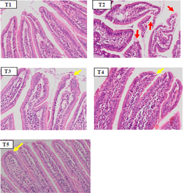
Figure 1
Hispathology of rat duodenum (Rattus norvegicus) with HE staining 100x magnification showed there was an erosion on duodenal villi epithelial (red arrow) in positive control group (T2) due to indometasin. The SHM group (T3,T4,T5) showed a improvement in duodenal villi epithelial damage.
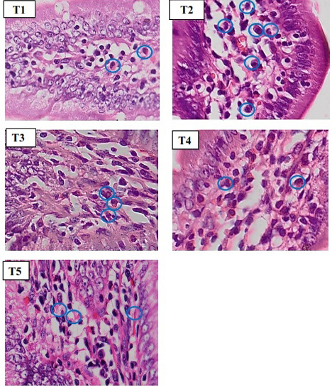
Figure 2
Histopathology of rat duodenum (Ratus morvegicus) (HE staining 400x magnification) revealed that group T2 contained more inflammatory cells (blue circle) than in all groups.


