Advances in Animal and Veterinary Sciences
Research Article
Adv. Anim. Vet. Sci. 9(9): 1400-1407
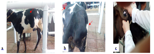
Figure 1
Notice distension of left side of the abdomen (a), Bilateral distension of the abdomen with left ventral and dorsal distension and right ventral distension (apple/pear) (b). Congested eye capillaries and sunken eye (c).
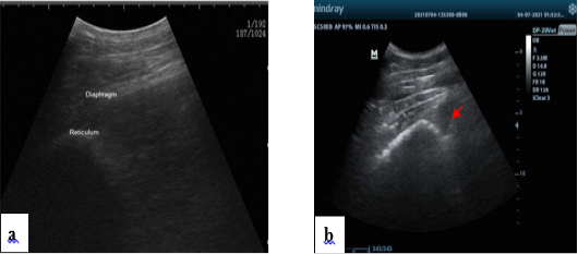
Figure 2
Normal ultrasonogram of the reticulum, notice half-moon shape (a). Presence of hypoechogenic fluid notice fibrin thread “arrow” in the reticular area in some diseased conditions (b).


