Advances in Animal and Veterinary Sciences
Research Article
Adv. Anim. Vet. Sci. 9(7): 1095-1112
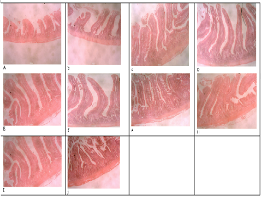
Figure 1
Histomicrograph of the duodenum of birds fed T1 (a), T2 (b), T3 (c), T4 (d), T5 (e), T6 (f), T7 (g), T8 (h), T9 (i), T10 (j), showing the villi height, crypt depth, thickness of the epithelium and that of the muscularis.
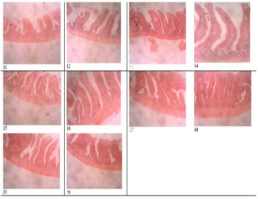
Figure 2
Histomicrograph of the jejunum of birds fed T1 (11), T2 (12), T3 (13), T4 (14), T5 (15), T6 (16), T7 (17), T8 (18), T9 (19), T10 (20), showing the villi height, crypt depth, thickness of the epithelium and that of the muscularis.
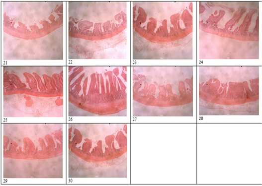
Figure 3
Histomicrograph of the ileum of birds fed T1 (21), T2 (22), T3 (23), T4 (24), T5 (25), T6 (26), T7 (27), T8 (28), T9 (29), T10 (30), showing the villi height, crypt depth, thickness of the epithelium and that of the muscularis.


