Advances in Animal and Veterinary Sciences
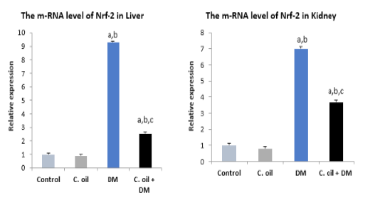
Real time PCR for Nrf-2 gene expression in liver and kidney of different experimental groups.
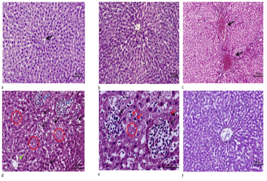
a. Liver tissue of control showing normal cell plates formed of polygonal hepatocytes with granular eosinophilic cytoplasm and vesicular nuclei & intervening regular sinusoids and normal central veins (black arrows). (H&E stain ×100). b. Liver of C. oil treated rat showing normal structure of the liver with normal central vein, sinusoids and hepatocytes (H&E stain ×100). c. Liver of DM treated rats showing marked portal veins congestion (black arrow) associated with widely diffuse hydropic degeneration of hepatocyte (H&E stain ×100). d. Liver from DM group showing diffuse marked hepatocellular hydropic degeneration with dilated hepatic sinusoids and multiple foci of hemorrhage (black arrows) with areas of necrosis showing ghost of hepatocytes some of hepatocytes revealed condensed/pyknotic nuclei (red circle) with aggregates of mononuclear inflammatory cells (blue arrows) ).(H&E stain ×200). e. High power of pervious image showing aggregates of mononuclear inflammatory cells (black arrows) surrounding necrotic hepatocytes & areas of ghost of cells and few hepatocytes with condensed/pyknotic nuclei (red arrows) (H&E stain ×400). f. Liver section from C. oil plus DM group showing normal hepatic architecture with regeneration of hepatocytes and improvement in central vein and sinusoidal blood (H&E stain ×100).
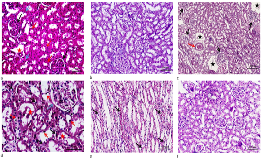
a. Renal section from control, showing normal glomeruli formed of capillary tuft surrounded by Bowman’s capsule ( black arrows), proximal convoluted tubules with narrow lumen lined by high cuboidal cells with homogeneous eosinophilic cytoplasm (red arrows) and distal convoluted tubules with wide lumen lined by low cuboidal cells (blue arrows) with normal- sized interstitial blood vessel (red star).(H&E stain ×200). b. Renal section from C. oil group, renal tissue demonstrated unremarkable histopathological changes. c. Renal section from DM group, cortical renal tissue demonstrated diffused moderate capillaries glomerular tuft congestion with Bowman’s capsule swelling (black arrows) note that the surrounding renal tubules showed degenerative changes with moderate interstitial edema (blue stars) and congestion (red arrow).(H&E stain ×100). d. Renal section from DM group, cortical renal tissue demonstrated diffused moderate congestion of glomerular tufts (blue arrow) & interstitial congestion (black arrows). The epithelial cells lining renal tubules showed diffuse hydropic degenerative swelling, noting some of cells had dark small pyknotic nuclei with few intraluminal sloughed cells (red stars).(H&E stain ×400). e. Renal section from DM group, medullary renal tissue demonstrated marked diffused interstitial edema and congestion with extravasation RBCS (black arrows), noting the diffuse degenertative changes of distal convoluted renal tubules (H&E stain ×200). f. Renal section from DM plus C. oil group, demonstrated mild hydropic degeneration of tubular cells and nearly normal glomeruli & interstitial tissue. (H&E stain ×200).


