Advances in Animal and Veterinary Sciences
Research Article

Gross Morphological Studies on the Air Sacs in Rosy-faced Parrots (Agapornis roseicollis)
Pamela Bejdić1*, Amela Katica1, Nadžida Mlaćo1, Lejla Velić2, Amel Ćutuk3, Benjamin Čengić4
1Department of Anatomy and Histology with Embryology, Veterinary Faculty, University of Sarajevo, Bosnia and Herzegovina; 2Department of Microbiology and Infectious diseases, Veterinary faculty, University of Sarajevo, Bosnia and Herzegovina; 3Department of Ambulatory Clinics, Veterinary Faculty, University of Sarajevo, Bosnia and Herzegovina; 4Department for Obstetrics and Udder diseases, Veterinary Faculty, University of Sarajevo, Bosnia and Herzegovina.
Abstract | The aim of this study was to morphologically describe the air sac system in Rosy-faced parrots (Agapornis roseicollis) and provide useful data for future veterinary care and treatment of respiratory system diseases in these pets. The research was conducted on five birds. In order to obtain the casts of the air sacs, we applied the corrosion cast technique, where we used the 26% solution of Vinylite mass. The research showed that the anatomy of the air sacs in these parrots was very similar to that in other birds, but there was some specific characteristic regarding the arrangement and connection between the air sacs. In Rosy-faced parrots we identified nine air sacs, the one unpaired, saccus clavicularis and paired saccus cervicalis, thoracicus cranialis, thoracicus caudalis and abdominalis. The casts showed that clavicular and cranial thoracic air sacs established a connection through the diverticula sternalia. These anatomical characteristics can be common to birds from Psittaciformes order and additional research need to be performed to confirm these findings.
Keywords | Rosy-faced lovebirds, Parrots, Air sacs, Anatomy
Received | March 12, 2021; Accepted | March 17, 2021; Published | June 01, 2021
*Correspondence | Pamela Bejdić, Department of Anatomy and Histology with Embryology, Veterinary Faculty, University of Sarajevo, Bosnia and Herzegovina; Email: pamela.bejdic@vfs.unsa.ba
Citation | Bejdić P, Katica A, Mlaćo N, Velić L, Ćutuk A, Čengić B (2021). Gross morphological studies on the air sacs in rosy-faced parrots (agapornis roseicollis). Adv. Anim. Vet. Sci. 9(7): 989-993.
DOI | http://dx.doi.org/10.17582/journal.aavs/2021/9.7.989.993
ISSN (Online) | 2307-8316; ISSN (Print) | 2309-3331
Copyright © 2021 Bejdić et al. This is an open access article distributed under the Creative Commons Attribution License, which permits unrestricted use, distribution, and reproduction in any medium, provided the original work is properly cited.
Introduction
The Rosy-faced (Agapornis roseicollis), also known as the Rosy-collared or Peach-faced lovebirds, are colourful, small in size, parrots from Psittaciformes order. These birds originated from south-west Africa, where they inhabit a dry, woodland, semi-desert and mountainous areas situated above 1600m of altitude (Radamaker and Corman, 2011). Due to their small size, beautiful colour of the feathers, easy maintenance and breeding, these parrots are often kept in captivity as pets. During the mid-1980s some of these birds escaped from the captivity and established healthy, breeding population (Andreasen et al., 1995) in the area of Phoenix and Arizona (USA). In veterinary practice is well known that birds kept in captivity are highly susceptible for different respiratory diseases. Recently there have been a couple of reports from different veterinary clinics regarding the treatment of respiratory system diseases in these parrots (Cooper et al., 2015; Andreasen et al., 1995; Nakano and Une, 2016). However, in available literature we did not find any data about the anatomy of respiratory system in these parrots. It is well known that the bird’s respiratory system is structurally one of the most complex and in terms of functionality, the most efficient among the vertebrates (Maina 2008; 2015). Its complex anatomical arrangement is a direct reflection of the adaptation of the birds to the flight and different habitats. The birds possess system of the air sacs and parabronchi connected to the lungs, and those structures represent them morphologically unique in the animal kingdom (Getty 1975; Nickel et al., 1977; King and McLelland 1984; Baumel et al., 1993; Dyce et al., 2002; König et al., 2016). In the past, many research have been conducted with the aim to morphologically describe and functionally explain the air-sacs system in different avian taxa (Kloek and Casler, 1972; Kadosaki 1977. and 1978; Hamlet and Fisher, 1967; Demirkan et al., 2006; Onuk et al., 2009; Orhan et al., 2009; Sawad and Udah 2012.; El-Bably et al., 2014; Casteleyn et al., 2010; Ragab and Reem 2016; Jaifar and Sawad 2006). The results of these research confirm that there are many differences among the birds’ species, particularly in the number, size and shape of the air sacs and their diverticula. In regard to all mentioned above, the main aim of this study was to morphologically describe the air sac system in Rosy-faced parrots and by doing so, provide useful data for future veterinary care and treatment of respiratory system diseases in these pets.
Materials and Methods
The research was conducted on five Rosy-faced lovebirds, two males and three females. All birds were aged from three to six years and did not have any health issues. The birds were kept in a private household as pets, where they died from freezing after the heating system collapsed. The owner donated the birds to the University of Sarajevo, the Veterinary faculty, the Department of Anatomy and Histology with Embryology, where we performed detailed morphological study of the lower part of their respiratory system, particularly the air sacs. For this examination, we applied the corrosion cast technique (Tompsett, 1970). The casts of the lower part of the respiratory system were prepared by using the 26% solution of Vinylite mass dissolved in acetone p.a. The prepared mass was additionally coloured in yellow (BIODUR® Colour Paste Corn Yellow AC 53) for better visualisation. The Vinylite mass was injected into the lumen of the trachea by plastic syringe with needle. During the injection, the pressure on abdominal wall was constantly monitored by manual palpation. When desired pressure was achieved, the injection of the Vinylite mass was stopped and the trachea was enclosed by surgical forceps. The birds were emerged into the tap water for 24 hours so that the Vinylite mass would harden. Later, three birds (two female and one male) ware placed into the device for soft tissue maceration of biological specimens (Schaerer, Weber-Bern), and the other were dissected. Dissected specimens were used for in situ morphological investigation and description of the lungs, air sacs and their diverticula. All dissected specimens were later permanently preserved in 3% formalin. The other birds were macerated and these specimens were later used for detail visualisation of the lungs and air sac casts. The process of maceration lasted 21 days. After that period, the specimens were washed in tap water and left to dry for 24 hours. At the end of this process, we got the full cast of the lungs and air sacs with preserved skeleton. For better visualisation of the casted structures we removed some of the bones. On these specimens we determined the position, shape and size of the air sacs and their diverticula. For proper Latin terminology we used the Nomina Anatomica Avium (Baumel et al., 1993).
Results
The lungs of Rosy-faced parrots are small and situated in the dorsal and lateral parts of the thoracic cavity. The five deep costal grooves can be identified on their costal and vertebral surface (Figure 1). The lungs covered the heart from above and caudally reached the cranial margin of the kidneys. The Vinilyte casts show that Rosy-faced parrots have nine air sacs and there is no difference between male and female birds. The one, saccus clavicularis was unpaired, while the other, saccus cervicalis, saccus thoracicus cranialis, saccus thoracicus caudalis and saccus abdominalis were paired (Figure 2, 3).
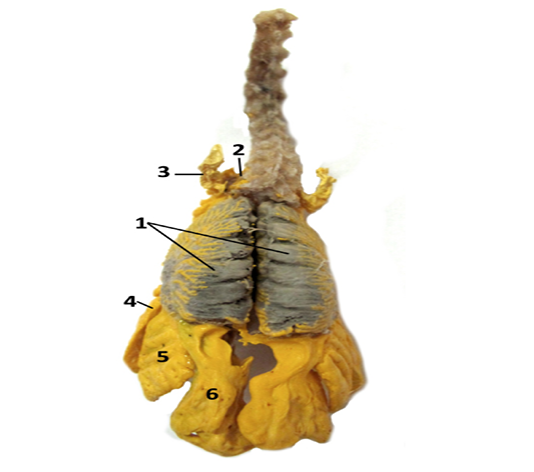
Figure 1: A photograph showing the dorsal view on the lungs and air sacs in Rosy-faced lovebirds dissected in situ: Photograph showing: 1. lungs, 2. cervical air sacs, 3. diverticulum suprahumerale of the clavicular air sac, 4. cranial thoracic air sac, 5. caudal thoracic air sac and 6. abdominal air sac.
The clavicular air sac occupied the cranial and central part of the thoracic cavity. Its main chamber lied between the pectoral girdle, sternum and the base of the heart (Figure 2). From its main chamber the six diverticula, two intra-thoracic and four extra-thoracic separated (Figure 1, 2). The intra-thoracic were diverticula cardiaca and sternalia. The cardiac diverticula covered the base of the heart and atrial wall. The sternal diverticula emerged from caudoventral part of the clavicular air sac. From its lateral side emerged one tubus-like extension through which this deverticula connected with the cranial thoracic air sac (Figure 2, 3). The four extra-thoracic diverticula of the clavicular air sac were: diverticulum subpectorale, suprahumerale, axillare and subscapulare (Figure 2, 3). The subpectoral diverticulum emerged from craniolateral side of the clavicular air sac. These diverticula had a triangular shape and laterally they are pierced by the axillar artery and vein. The left and right subpectoral diverticula were connected by one large extension which was situated above the trachea (Figure 4). Small axillary diverticula emerged laterally from the previous. It has a leaf-like shape and occupied the ventrolateral side of the shoulder joint. The suprahumeral diverticula were much larger and emerge above the axillary diverticula (Figure 1, 2, 3). They covered the head of the humerus. The subscapular diverticula is the smallest of all and occupied the spaces between the first two thoracic vertebrae (Figure 3).
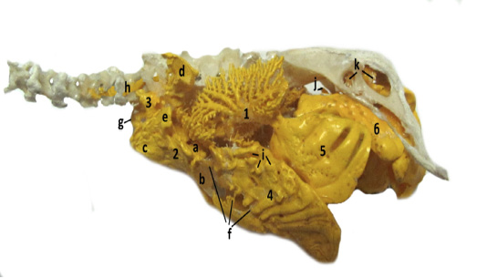
Figure 2: A photograph of lateral, in situ view of corroded and macerated bronchi and air sacs system showing: 1. lungs, 2. clavicular air sac with its diverticula cardiaca (a), sternalia (b), subpectorale (c), suprahumerale (d) and axillare (e), 3. cervical air sac with its diverticulum intramusculare (g) and vertebrale (h), 4. cranial thoracic air sac with its connections with clavicular air sac (f) and small dorsal diverticula (i), 5. caudal thoracic air sac, 6. abdominal air sac with its divertiucula perirenalia (j) and femoralia (k).
The cervical air sacs were small, paired and situated between the subpectoral diverticula of the clavicular air sac (Figure 4). These sacs gave two diverticula (diverticulum vertebralia and intermuscularia) that followed the vertebral column from the first thoracic to last five or six cervical vertebrae. The vertebral diverticula entered into the transversal and vertebral canal while the intermuscular diverticula penetrated the muscles in the caudal neck region (Figure 2, 3).
The cranial thoracic air sacs were large, oval and situated on the sternal parts of the ribs. Their dorsal margin reaches the point where vertebral and sternal extremity of the ribs joined. Only small part of these sacs protruded caudally from the rib cage. From their lateral and cranial side emerged small diverticula which aerates the vertebral part of the ribs. Similar diverticula exited from their ventral margin and aerate the first six sternal ribs (Figure 2, 3). The cranial thoracic air sacs connect with the clavicular air sac through their diverticula sternalia as it described above (Figure 2, 3).

Figure 3: A photograph of lateral, in situ view on corroded and dissected specimen where are visible: 1. lungs, 2. clavicular air sac with its diverticula cardiaca (a), sternalia (b), subpectorale (c), suprahumerale (d) and axillare (e), subscapulare (l), 3. cervical air sac with its diverticulum intramusculare (g) and vertebrale (h), 4. cranial thoracic air sac with its small dorsal diverticula (i), 5. caudal thoracic air sac, 6. abdominal air sac, 7 trachea, 8. heart.
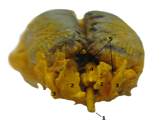
Figure 4: A photograph of cranial view on corroded and in situ dissected specimen where are visible: 1. trachea, 2. cervical air sacs, 3. diverticulum subpectorale of clavicular air sac and theirs connection (4).
The caudal thoracic air sacs were smaller than the cranial and they were situated on the vertebral parts of the ribs (Figure 2). The small, caudodorsal part of these sacs protrudes caudally from the last pair of the ribs. These air sacs did not give any diverticula or aerate the bones.
The abdominal air sacs were the largest and most voluminous. They occupied the dorsolateral side of coelomic cavity, extending from the caudal margin of the lungs to the cloaca (Figure 2). The right abdominal sac was slightly larger than the left. In the middle part, the left abdominal sac was constricted (Figure 1). These sacs gave small perirenal and femoral diverticula (Figure 2). The perirenal diverticula emerged from their dorsolateral side and aerate the synsacrum and osilium. The femoral diverticula separate at the level of the hip joint and penetrate the acetabulum, ilium and ishium.
Discussion
The anatomy of the lower part of the respiratory system is quite variable among the avian taxa and these variations come as results of bird’s adaptation to the flight and different habitats (Maina, 2015). The most intensive research on the air sac system so far has been conducted on domestic birds. In today’s official literature it has been stated that domestic Fowls have eight air sacs, an unpaired cervical and clavicular sack and paired cranial thoracic, caudal thoracic and abdominal air sacs (King and McLelland 1984; Baumel et al., 1993; Dyce et al., 2002; König et al., 2016). However, in some reports it has been noted that there are nine air sacs present in these birds (Maina, 2008). These differences appear as results of a different description of the cervical air sac. It is well known that the cervical air sac in domestic fowl are paired during the embryonic development, while later, after the hatching they fuse and form one main chamber from which two thin diverticula separate and goes bilaterally along the cervical vertebra (King and McLelland, 1984; Baumel et al., 1993). In some research these diverticula have been described as paired cervical air sacs, which leads to a different interpretation of the number of air sacs (Maina, 2008). The similar anatomical arrangement of the air sacs also has been found in some wild birds such as Mallard and Golden Peking Duck (Demirkan et al., 2006), Long-legged Buzzard (Orhan et al., 2009), Bulbul (Jaifar and Sawad, 2016), some birds species from Procellariiformes order (Hamlet and Fisher, 1967), Strigidae and Ardeidae family (Kadosaki, 1977), while in some species it has been recorded the fusion of the cervical and clavicular air sacs, and that leads to their lesser number. The fusion of the air sacs have been found in Japanese Quails (Sawad and Udah, 2012), Turkey (Ragab and Reem 2016), Goose (Onuk et al., 2009) and Pigeon (Nickel et al., 1977). However, in present literature we did not find any data about the anatomy of the respiratory system in Rosy-faced lovebirds. Our research showed that the lungs and the air sacs system in these parrots were very similar to those in domestic fowls, but there are some important differences among these two species. In Rosy-faced lovebirds, the cervical air sacs were paired, but they had the similar diverticula as in fowls. However, the cervical diverticula did not extend cranially to the axis as in fowl. They extended only from the first two thoracics to the last five or six cervical vertebrae. The clavicular air sac in our birds was unpaired as in Fowls, but there are some significant differences among the species. In these birds, the clavicular air sac was connected through their diverticula sternalia with cranial thoracic air sacs. The connections between these sacs so far have been recorded in a Common grackle, Cardinals, Rufous-sided Towbees, Song Sparrow and some individual Old World song birds (Kloek and Casler, 1972), but not in fowls or other domestic birds (King and McLelland, 1984; Baumel et al., 1993; Dyce et al., 2002). Also, in Rosy-faced lovebirds we identified a connection between the left and right subpectoral diverticula of the clavicular air sac, which was not identified in other birds species. In domestic fowls and the bulbul, it has been found that the cranial thoracis air sacs are smaller than the caudal and both of them do not possess any diverticula (Nickel et al., 1977; King and McLelland 1984; Dyce et al., 2002; Onuk et al., 2009). This result was partially similar to the current findings. It was found that cranial thoracic air sacs in Rosy-faced lovebirds were larger then caudal and from their ventral margin and cranio lateral side emerged small diverticula which aerate the ribs. Similar observations have been recorded in Long-legged Buzzard (Onuk et al., 2009), Golden Peking (Onuk et al., 2009) and Mallard Duck (El-Bably et al., 2014). Additionally, in Long-legged Buzzard two specific diverticula of the cranial thoracic sacs have been recorded, diverticulum subcardiale and supracardiale, which was absent in the parrots (El-Bably et al., 2014). The caudal thoracic air sac did not have any diverticula and that is in agreement with the previuos research (King and McLelland 1984; Baumel et al., 1993; Dyce et al., 2002). Also, as in many other birds, it was recorded that the abdominal air sacs were the largest and most voluminous. In domestic fowls, the right abdominal sac is slightly larger than the left and our research coincides with these findings, with the addition that on the left sac a small constriction in its middle part was identified (Nickel et al., 1977; König et al., 2016). In conclusion, the present research showed that the Rosy-faced lovebirds have a specific anatomical arrangement and connection between the air sacs and additional research needs to be carried out to see are these features generally common to the birds from Psittaciformes order.
acknowledgements
The authors are grateful to Jasmina Mulaosmanović for the professional English review and editing.
Conflict of interest
The authors have declared no conflict of interest.
Author’s contribution
Pamela Bejdić proposed the study design, participated in necropsy and corrosive cast interpretation. Lejla Velić, Amel Ćutuk and Benjamin Čengić were involved in sample collection and corrosive cast preparation and maceration. Amela Katica and Nadžida Mlaćo participate in data revision and manuscript preparation. All authors have contributed, reviewed and approved the last submitted version.
References


