Advances in Animal and Veterinary Sciences
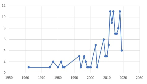
The count of research studies directed toward “Dermatomycoses in pets” through the period between 1962 and 2019.
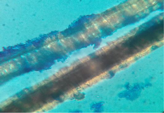
Dog hair sample stained with Lactophenol cotton blue examined microscopically (40X) showing ectothrix hair invasion with small spores.
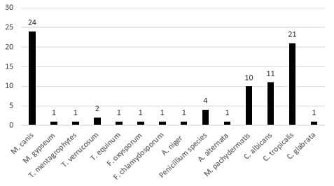
Clustered bar chart for the number of each fungal isolate.
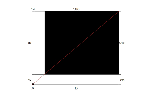
The agreement correlation of the two conventional laboratory testing methods used during the study, microscopic examination, and culture.
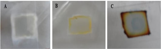
Macroscopic growth on SDA agar blocks of microculture set.
A for T. mentagrophytes; B for M. canis and C for T. equinum.
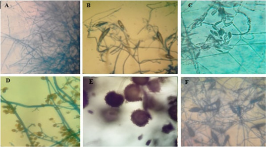
Microscopic pictures (using 40x high dry power lens) of different characteristic structures obtained from microculture technique.
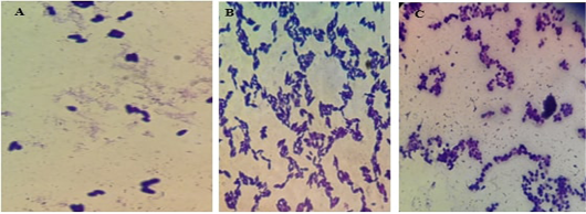
Microscopic pictures using the oil immersion lens of different yeast isolates stained with gram’s technique.
A for M. pachydermatis showing the characteristic bowling pins pattern, B for C. tropicalis and C for C. albicans.


