Advances in Animal and Veterinary Sciences
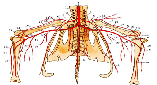
Arteries of the pelvis, thigh and leg regions, ventral view; diagrammatic
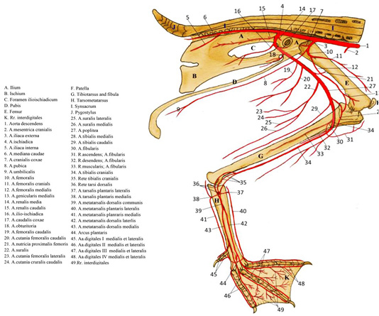
Arteries of the left pelvic limb of goose, medial view; diagrammatic.
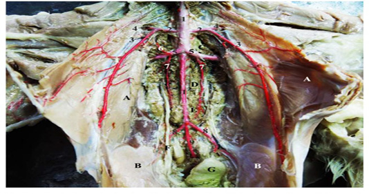
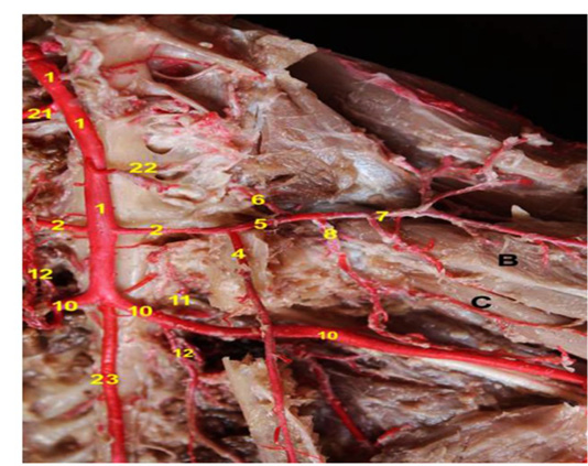
A photograph showing the termination of the descending aorta and the main arteries of the pelvis, (Ventral view).
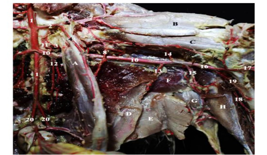
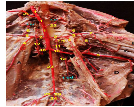
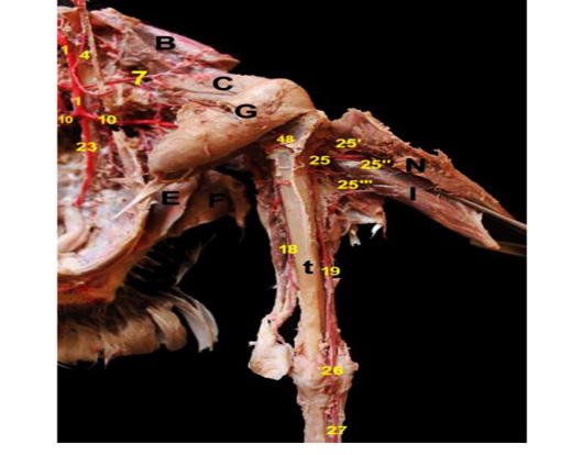
A photograph showing the arteries of pelvic and thigh regions; (ventro-medial view); left side.
A photograph showing the arteries of thigh region. (Ventro-medial view).
A photograph showing the arteries of pelvic and thigh regions. (Ventro-medial view)
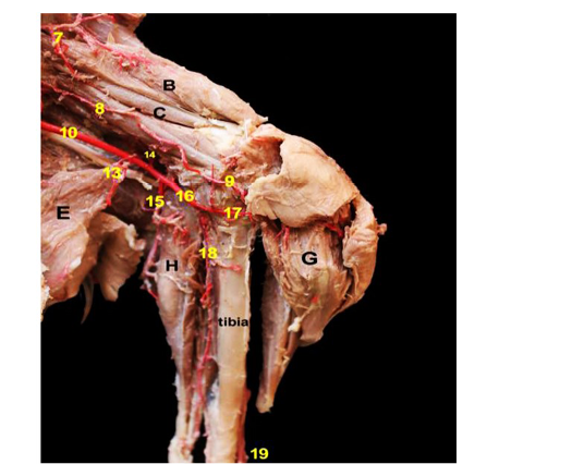
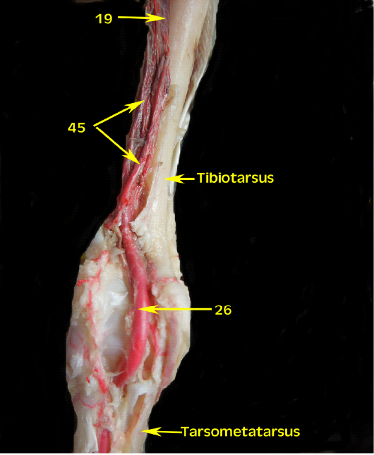
A photograph showing the arteries of the thigh and leg regions. (Caudo-medial view).
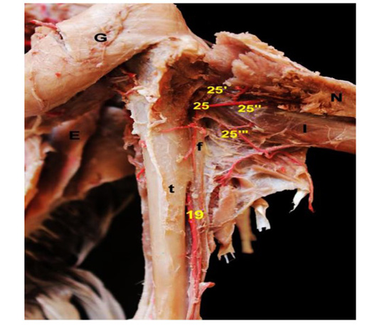
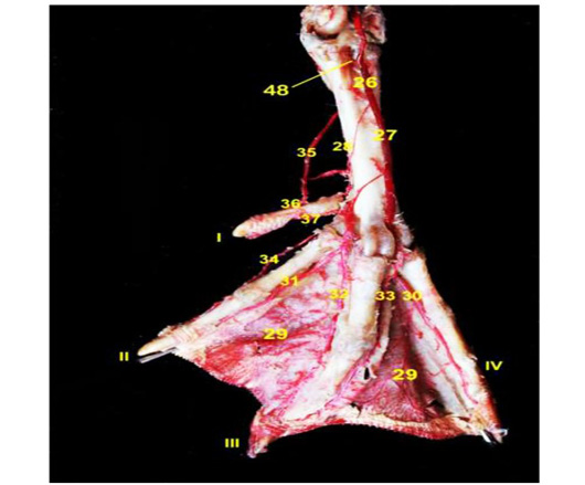
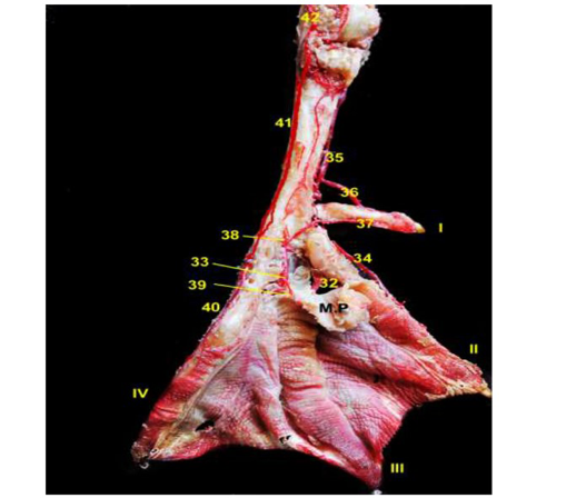
A photograph showing the branches of Fibular artery. (Dorso-lateral view)
A photograph showing the branches and continuation of the fibular artery (Dorso-lateral view).
A photograph showing the Rete tibiotarsale. (dorsal view)
A photograph showing the distribution of the digital arteries, dorsal view.
A photograph showing the distribution of the plantar arch, plantar view.


