Advances in Animal and Veterinary Sciences
Short Communication
Adv. Anim. Vet. Sci. 9(3): 438-441
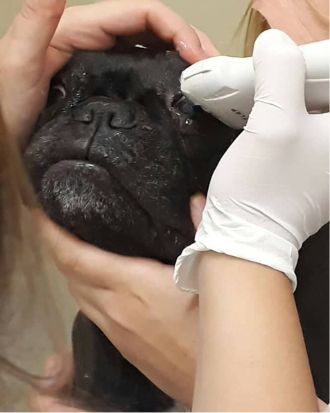
Figure 1
Ocular US evaluation in a female French Bulldog. Delicately contained in sitting position. Blepharostase was manual, and the exam was performed after instillation of 1 drop of anesthetic eye drops and sterile gel was used as a means of contact, using the transcorneal method.
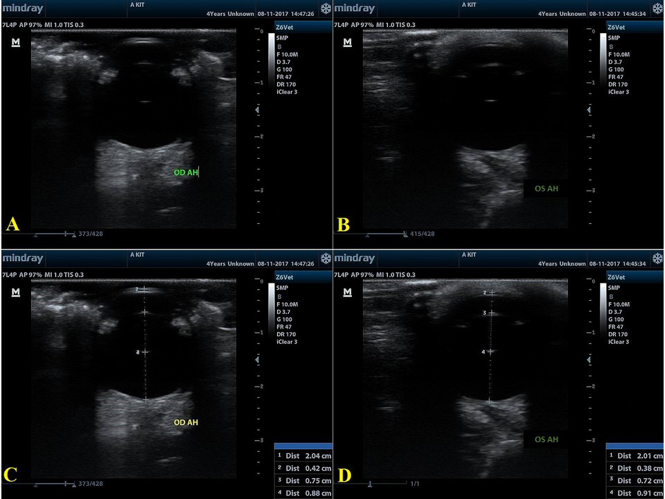
Figure 2
Representative B-scan ultrasonogram of female French Bulldog (A) and (B) representative B-scan ultrasonogram after optimal positioning was achieved and in (C) and (D) after the measures were done. Measurement values: Right eye (OD): (C) HAD = 2.04 cm (20.4 mm); ACD= 0.42 cm (4.2 mm); LT= 0.75 cm (7.5 mm); VCD= 0.88 cm (8.8 mm). Left eye (OS): (D) HAD= 2.01 cm (20.1 mm); ACD= 0.38 cm (3.8 mm); Lens Thickness= 0.72 cm (7.2 mm); VCD= 0.91 cm (9.1 mm).


