Advances in Animal and Veterinary Sciences
Case Report
Advances in Animal and Veterinary Sciences. 1 (2S): 24 – 25Special Issue–2 (Clinical Veterinary Practice– Trends)
Unilateral wing Amputation for the Management of Humerus Fracture in a Black Kite (Milvus Migrans)
Madhu Doddhadasarahalli Nanjappa, Shongsir Warson Monsang, Hari Prasad Aithal, Amarpal, Abhijit Motiram Pawde, Prakash Kinjavedkar, Malik Mohammed Shamsuz Zama
-
Indian Veterinary Research Institute (IVRI), Izatnagar, Uttar Pradesh, India
*Corresponding author:madhu63vsr@gmail.com
ARTICLE CITATION:
Nanjappa MD, Monsang SW, Aithal HP, Amarpal, Pawde AM, Kinjavedkar P and Zama MMS (2013). Unilateral wing amputation for the management of humerus fracture in a black kite (Milvus migrans). Adv. Anim. Vet. Sci. 1 (2S): 24 – 25.
Received: 2013–12–04, Revised: 2013–12–25, Accepted: 2013–12–27
The electronic version of this article is the complete one and can be found online at
(
http://nexusacademicpublishers.com/table_contents_detail/4/169/html
)
which permits unrestricted use, distribution, and reproduction in any medium, provided the original work is properly cited
ABSTRACT
An adult common Pariah kite was presented with the history of trauma in right wing. Physical and radiographic examination revealed fracture of distal third of right humerus. The exposed bones were necrosed along with the loss of surrounding soft tissues. Amputation of wing at proximal third of humerus was done under general anesthesia to save the life of the bird. Proper postoperative care and management ensured the bird to recover from the loss of wing by thirty days postoperatively without any complications.
The black kite or common pariah kite (Milvus migrans) is a medium–sized bird of prey in the family Accipitridae, which also includes many other diurnal raptors. It is thought to be the world's most abundant species of acciptrid, although some populations have experienced dramatic declines or fluctuations. The common Pariah kite, Milvus migrans govinda, a race of the black kite, which in various forms has a very wide distribution in the old world, is found throughout India, Burma and Ceylon, extending still farther east to Hainan. Bone fractures are common in both wild and captive birds (Fix and Barrows, 1990; Houston, 1993). Avian bones are thin and brittle and tend to break into fragments upon a variety of natural events like midair collisions, fights with other birds (Houston, 1993) or anthropogenic experiences like gunshot wounds, collisions with automobiles or fences, encounters with traps, attacks by dogs or cats, etc. (Fix and Barrows, 1990).
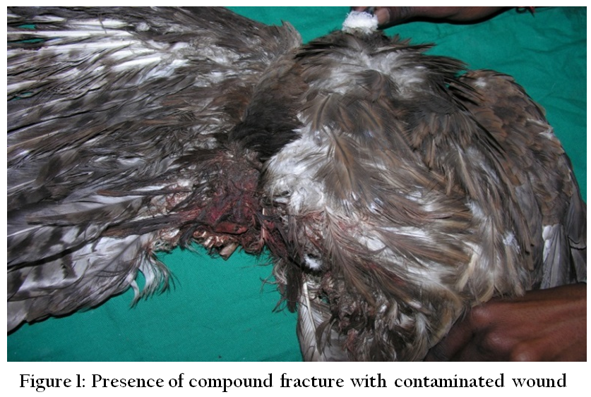
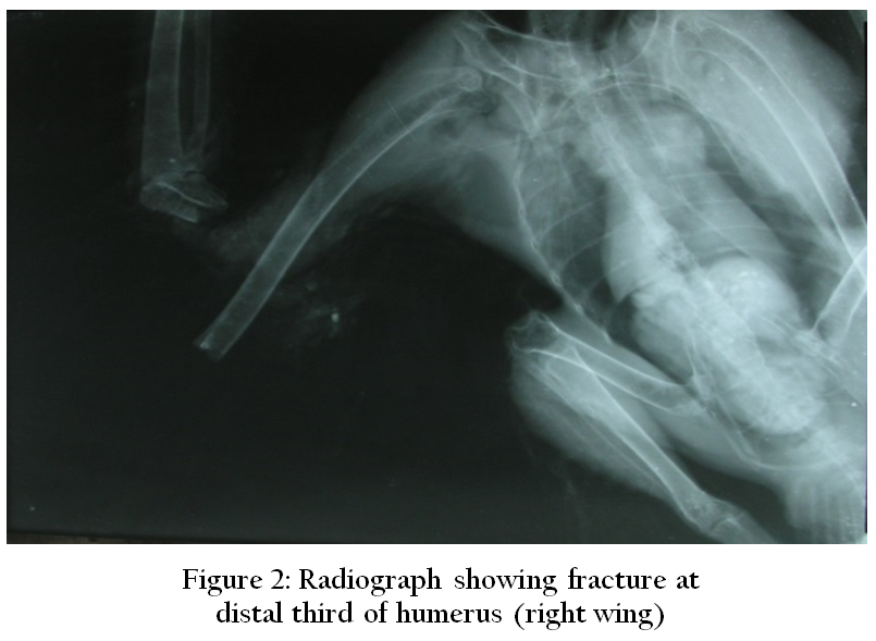
An adult, common pariah kite weighing 2.5 kg was rescued by the forest officials from the outskirts of Bareilly city and brought to the Referral Veterinary Polyclinics, IVRI for treatment of wound in right wing. Physical examination revealed the bird to be dull and depressed with exposed bones of right wing having sharp edges along with loss of soft tissue coverage. The fractured fragments were necrosed (Figure 1) with the presence of contaminated wound on the medial aspect of distal third of the humerus. However, the contralateral wing appeared nearly normal and rest of the body did not reveal any abnormality or injury. Radiographic examination confirmed the fracture of distal third of humerus of the right wing (Figure 2). Based on the clinical and radiographic findings, amputation of the wing was considered.
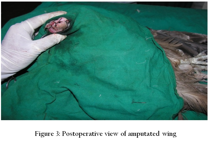
The bird was administered with xylazine (Xylaxin®, Indian Immunologicals Ltd; 5 mg/kg, IM) followed 10 minutes later by ketamine (Aneket®, Neon Labs; 15 mg/kg IM). The bird was restrained in left lateral recumbency and the surgical site was prepared for aseptic surgery by plucking the feathers. The wounded site was flushed with lukewarm normal saline mixed with 1% chlorhexidine solution and the adjoining feathers were plucked off around the injury site to prevent further wound infection. The wound was then thoroughly debrided followed by topical application of 5% povidone iodine solution. The wing was prepared and draped for aseptic surgery in a routine manner. The skin was incised and muscles were transected. A transverse osteotomy was carried out at the proximal third of the humerus after raising the muscle flaps from a healthy area. The muscles were sutured using chromic catgut No. 2–0 over the bone stump. The subcutaneous tissue was sutured using chromic catgut No. 2–0. Finally the skin was closed by black braided silk No. 2–0 (Figure 3). The owner was advised to keep the bird confined in a cage to restrict movement for a minimum period of two weeks.
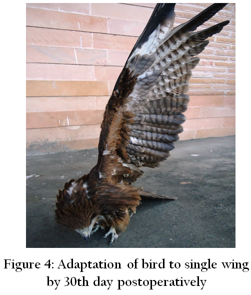
Postoperatively, meloxicam (Melonex®, Intas; 0.5 mg/kg IM, once daily for 5 days) and enrofloxacin (Bayrocin®, Pfizer–Bayer; 20 mg/kg IM, once daily for 7 days) were administered along with antiseptic wound dressing for 10 days. The skin sutures were removed on 10th day postoperatively. The bird recovered completely without any complications and became completely normal by 30 days (Figure 4). However, the bird lost its functional ability to take a long flight and could only use its wings for very small distance flights.
Most avian patients are under severe stress after a fracture because of the initial trauma and the additional stress of restraint and handling (Withrow, 1982). In many cases, these fracture sites are grossly contaminated and the fragments are necrosed. In such cases, fragment stabilization with standard orthopedic techniques may not be useful. Hence, to save the life of the wild birds, amputation of the necrosed bony fragments may be the only choice (Vineet Kumar, 2012). Avian bone fractures are often open and frequently comminuted, especially in wild birds (Bennett and Kuzma, 1992). A number of standard orthopaedic techniques have been used for fracture management in eagle by several scientific workers with variable results (Langley–Hobbs and Friend, 2002; Davidson et al., 2005; Guzman et al., 2007; Manjulkar et al., 2008). In the present case, necrosis and infection were seen on presentation of the bird. Therefore, amputation of the humerus at its proximal third was done in an attempt to prevent osteomyelitis and further spread of infection to other bones.
REFERENCES
Bennett RA and Kuzma AB (1992). Fracture management in birds. J. Zoo Wildlife Med. 23(1): 5–38.
Davidson JR, Mitchell MA and Ramirez S (2005). Plate fixation of a coracoid fracture in a Bald Eagle (Haliaeetus leucocephalus). J. Avian Med. Surg. 19: 303–308.
http://dx.doi.org/10.1647/2004-037.1
Fix AS and Barrows SZ (1990). Raptors rehabilitated in Iowa during 1986 and 1987: A retrospective study. J. Wildlife Dis. 26: 18–21.
http://dx.doi.org/10.7589/0090-3558-26.1.18
PMid:2304198
Guzman DS, Bubenik LJ, Lauer SK, Vasanjee S and Mitchell MA (2007). Repair of a coracoid luxation and a tibiotarsal fracture in a Bald Eagle (Haliaeetus leucocephalus). J. Avian Med. Surg. 21: 188–195.
http://dx.doi.org/10.1647/1082-6742(2007)21[188:ROACLA]2.0.CO;2
Houston DC (1993). The incidence of healed fractures to wing bones of White–backed and Ruppell's Griffon vultures Gyps africanus and G. rueppellii and other birds. Int. J. Avian Sci. 135: 468–475.
Langley–Hobbs SJ and Friend E (2002). Interlocking nail repair of fractured femur in a turkey. Vet. Rec. 150: 247–248.
http://dx.doi.org/10.1136/vr.150.8.247
PMid:11916027
Manjulkar GP, Zade PR and Pathak VP (2008). Use of PVC sheet for repair of fracture in eagle. Vet. World. 1: 119.
Vineet Kumar, Mathew DD, Rekha Pathak, Ahmad RA and Zama MMS (2012). Surgical management of an Indian spotted eagle with compound fracture of humerus. J. Adv. Vet. Res. 2: 301–302
Withrow SJ (1982). General principles of fracture repair in raptors. Compend. Cont. Edu. Pract. Vet. 4: 116–121.


