Advances in Animal and Veterinary Sciences
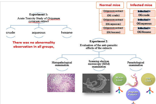
The experimental design of the two experiments
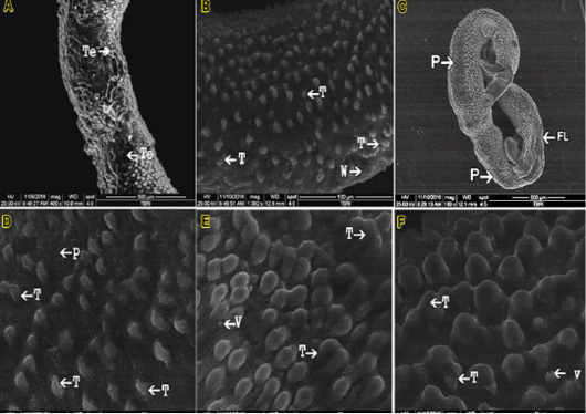
SEM micrograph (A) male S. mansoni after exposure to the crude Origanum syriacum; (B, C, D &E) male S. mansoni after exposure to the aqueous fraction of O.syriacum extracts; (F): male S. mansoni after exposure to the hexane fraction of O.syriacum extract
Tubercles (T); Wrinkling (W); Peeling (P) Vesicles (V); Focal lesions (FL).
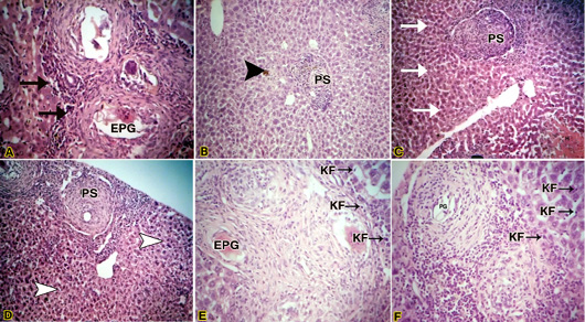
(A) Light micrograph of liver of infected mouse with S. mansoni treated with crude Origanum syriacum extract H&E. Magnification (400x); (B): Light micrograph of liver of infected mouse with S. mansoni treated with crude O.syriacum extract Magnification (100x); (C & D) Light micrograph of liver of infected mouse with S. mansoni treated with aqueous fraction of O.syriacum extract H&E. Magnification (100x); (E&F): Light micrograph of liver of infected mouse with S. mansoni treated with hexane fraction of O.syriacum extract H&E. Magnification (200x). Productive granuloma (PG); Exudative-productive granuloma (EPG); Kupffer cells (KF).


