Advances in Animal and Veterinary Sciences
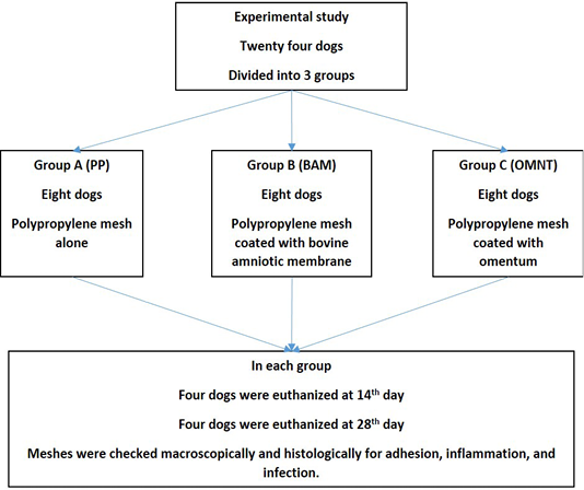
The experimental work design and groups identification.
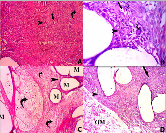
A photomicrograph of all groups at 14 days post-surgery. (A) Group A, showing PP mesh surrounded by marked infiltration of mononuclear inflammatory cells (score 4) (head arrow) with mild neovascularization (score 2) (straight arrow) and mild fibrosis (score 2) (Curved arrow), H and E, stain. X 100. (B) Foreign body giant multinucleated cell (score 1) (curved arrow), H and E, stain. X 400. (C) Group B, showing mesh (M) surrounded by minimal infiltration of mononuclear inflammatory cells (score 1) (head arrow) with persistence of vital AM (curved arrow) H and E, stain. X 100. (D) Group C, showing mesh surrounded by moderate infiltration of mononuclear inflammatory cells (score 3) (head arrow) with mild fibrosis (score 2) (straight arrow) and persistence of omentum (OM) next to the mesh (score 2), H and E, stain. X 100.
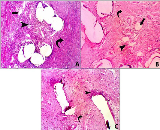
A photomicrograph of H and E stained sections for all groups at 28 days post-surgery. Group A, showing mesh surrounded by moderate infiltration of mononuclear inflammatory cells (score 3) (head arrow) with moderate fibrosis (score 3) (curved arrow) and minimal neovascularization (score 1) (straight arrow) X 100. Group B, showing mesh surrounded by mild infiltration of mononuclear inflammatory cells (score 2) (head arrow) with moderate fibrosis (score 3) (curved arrow) and minimal neovascularization (score 1) (straight arrow), X 100. Group C, showed mesh surrounded by mild infiltration of mononuclear inflammatory cells (score 2) (head arrow) with marked fibrosis (score 4) (curved arrow) X 100.
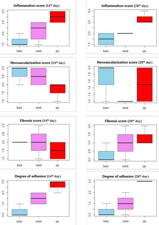
Score of inflammation, neovascularization, and fibrosis and degree of adhesion in all experimental groups at 14th and 28th days post-surgery according to a semi-quantative grading scale described by Deeken and Mathews (2012).


