Advances in Animal and Veterinary Sciences
Case Report
Advances in Animal and Veterinary Sciences. 2 (1): 46 – 49Gangrenous Mastitis: An Important Staphylococcus aureus Related Problem in Goat Husbandry
Arslan Tariq1*, Asim Shahzad2, Razia kuasar3, Asher Mehfooz4, Asad Manzoor4, Misbah Ijaz4, Imaad Rashid4, Nasir Mahmood1, Jahanzeb Tahir1, Syed Hamza Turab Zaidi1, Uzair Ahasan Fiaz1, Muhammad Salman Qureshi5
2. Department of Veterinary Pathology, Faculty of Veterinary Sciences (FVS), University of Agriculture Faisalabad (UAF), Pakistan.
3. Department of Veterinary Anatomy and Histology, Faculty of Veterinary Sciences (FVS), University of Agriculture Faisalabad (UAF), Pakistan.
4. Department of Clinics, Medicine and Surgery (CMS), Faculty of Veterinary Sciences (FVS), University of Agriculture Faisalabad (UAF), Pakistan.
5. Faculty of Veterinary Sciences (FVS), Gomal University Dara Ismail Khan, Pakistan.
*Corresponding author: dr.arslantariq3418@live.com
ARTICLE CITATION: Tariq A, Shahzad A, kuasar R, Mehfooz A, Manzoor A, Ijaz M, Rashid I, Mahmood N, Tahir J, Zaidi SHT, Fiaz UA and Qureshi MS (2014). Gangrenous mastitis; an important staphylococcus aureus related problem in goat husbandry. Adv. Anim. Vet. Sci. 2 (1): 46 – 49.
Received:2013–12–17, Revised:2013–12–25, Accepted: 2013–12–26
The electronic version of this article is the complete one and can be found online at (http://dx.doi.org/10.14737/journal.aavs/2014.2.1.46.49) which permits unrestricted use, distribution, and reproduction in any medium, provided the original work is properly cited
ABSTRACT
Inflammation, bluish discoloration of udder and teat skin; and increase in somatic cell count (SCC) are the main identification marks for gangrenous mastitis. Staphylococcus aureus is well known to produce gangrenous mastitis in milking animal’s especially small ruminants of 2–3 year of age. A typical case of gangrenous mastitis with characteristic signs and symptoms was recorded at department of Clinical, Medicine and Surgery (CMS), University of Agriculture, Faisalabad (UAF), Pakistan. Right teat of animal was almost necrosed which became sloughed–off on palpation leaving an open infected wound. Somatic cell count (SCC)>1x10⁶, pus in milk from left teat and round cream colonies of S. aureus on Staphylococcus 110 medium in bacterial culture provide base for tentative diagnosis as gangrenous mastitis. But confirmation occur after getting catalase positive (+) and coagulase positive (+ve) tests at 4 hours. Wound dressing with antiseptic, cover antibiotic and anti–inflammatory was recommended for 7 days along with pre and post teat dipping. Animal was on the way of recovery when checked after 12 days of treatment but animal lost one teat permanently.
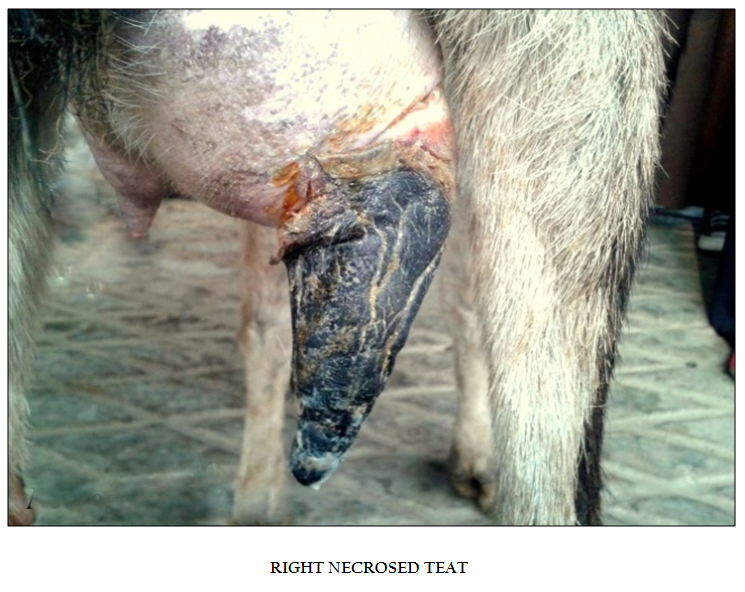
Mastitis reflects the inflammation of the mammary gland, which may occur due to any bacterial infection secondary to teat injury or poor management (Marogna et al., 2012; Egwu et al., 2001). Important causative agents for mastitis include Streptococcus spp., non–hemolytic Staphylococcus spp., Staphylococcus aureus, Pseudomonas spp. and Escherichia coli etc. Above listed organisms reported to produce 38.2%, 11.0%, 4.1%, 1.6% and 1.2% mastitis respectively (White and Hinckley, 1999). Among all of above, most important is Staphylococcus aureus in goat mammary gland system, can produce any type of mastitis like subclinical, chronic and acute. Increase pathogenic potential of S. aureus produces disease in more severe form i.e. gangrenous mastitis (White and Hinckley, 1999). High morbidity and mortality was observed along with 4% to 40% frequency of S. aureus among all isolated microorganisms recorded (Marogna et al., 2012). Clinically disease is characterized by inflamed udder, reduction and change in appearance and composition of milk. These signs are not seen in sub–clinical infection except increase in SCC and may develop into chronic mastitis. Factors influences the prevalence of mastitis in goats includes number of birth and lactation stage (Marogna et al., 2012). Egwu et al., (2001) mentioned more mastitis cases in 2–3 year old does because of increase in milk production in subsequent lactation. Diagnose of mastitis possible other than clinical examination of udder, by using other tests like California Mastitis Test (CMT), surf field mastitis test (SFMT) and Electrical Impedance test (EIT) etc. but still best standard is SCC(Marogna et al., 2012). Dairy industry mainly concern with food safety because of usage of goat milk as raw milk and for consumption (White and Hinckley, 1999). Rich supplementation of milk making the goat husbandry more popular, but contrary facing the problem of Mastitis, cause huge economic losses (Egwu et al., 2001; Marogna et al., 2012). According to Margona et al., (2012) 18% of animals are cull or die due to this disease. 40% prevalence of mastitis was reported at study area by Ali, (2008).Target of this study is the control of mastitis by maintaining hygiene environment and post–milking dipping. Beetal goat of approximately 3 years of age was reported to outdoor clinic at Department of Clinics Medicine and Surgery (CMS), University of Agriculture, Faisalabad (UAF), Pakistan with chief complaint of pain at right teat which became necrosed, almost black in color and cold to touch (Figure 1). Investigation revealed that the problem started with swelling of respective teat after 8day of kidding. Later on, on 14th day respective teat start darkening and a start secreting bloody
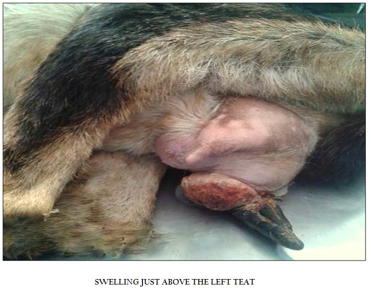
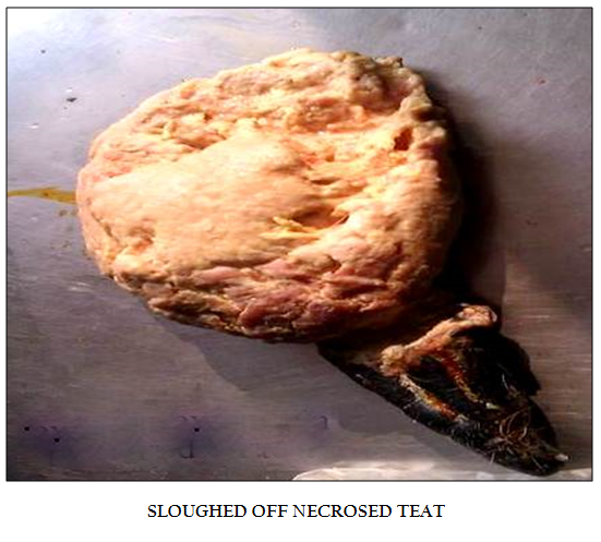
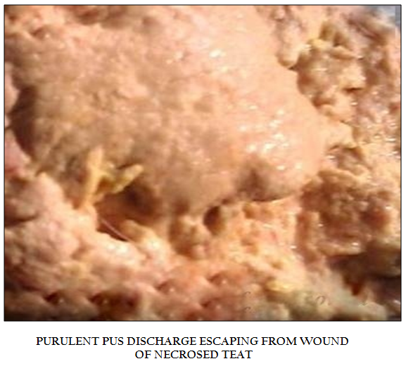
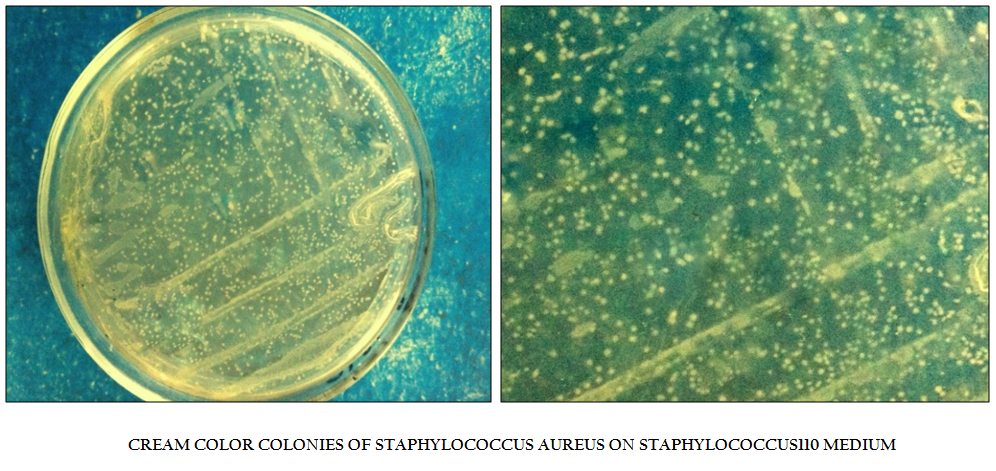
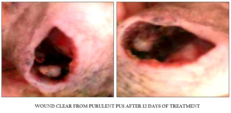
discharge containing pus. No treatment was done by the owner except massage of respective teat and udder with lukewarm salt water for sake of reduces swelling but all in vain and left a dark gangrenous dried mass. Case presented on 20th day of kidding. Clinically high fever (104.2), increase in heart beat (110 beats/min) was noted. Affected teat sloughed off during physical manipulation escaping a purulent exudate from acini and inter–lobular spaces (Figure 3~4) of udder also reported by Yeshwantkumar and Nirmala (2008). Left teat was normal apparently but a little swelling just above on udder was seen (Figure 2). Surf field mastitis test was performed for milk sample from left teat and found to be 2 positive (++) for mastitis with> 1x10⁶ Somatic cell count (SCC). Bacterial culturing of milk was done for identification of causative organism. This was done by taking milk sample with sterilizing loop and spread on the Staphylococcus 110 medium and incubated at 370C for 72 hours. Cream color colonies and of S. aureus were obtained on Staphylococcus 110 medium (Figure 5~6). After getting colonies; catalase and coagulase test were done. Catalase test was performed with 3% soln. of H2O2.of which 2 drops taken on slide and a loop full of culture is mixed in it; formation of bubbles was providing clue for presence of staphylococcus in culture. Coagulase test was done with rabbit plasma in which loop of culture was mixed and incubated at 370C and checked every hour for coagulation of plasma. Coagulation of rabbit plasma occurs at 4 hour. Positive (+) catalase test and positive (+) coagulase test on 4 hour confirmed the organism as S. aureus (Rashid, 2011), which is the main culprit in gangrenous mastitis. Clinical signs and symptoms increase in somatic cell count (SCC) and bacterial culture confirmed the disease as gangrenous mastitis. The sloughed off region was debrided with dilute solution of KMnO4and dressed with povidone iodine solution. Front–line spray (0.25% w/v fipronil) was recommended to spray on the dressed part every time after changing the bandage to protect the effected part from parasites or maggots. Treatment is usually done with NSAIDs and antibiotics but in most severe cases teat amputation is recommended (Ribeiro et al., 2007; Yeshwantkumar and Nirmala, 2008).About 91.67 % Susceptibility of mastitis causing organism against enrofloxacin is seen by Ali, (2008). So symptomatic treatment was done with Meloxicam @0.6mg/kg body weight intravenously and enrofloxacin@ 10mg/kg body weight subcutaneously for 7 days. Wound healing start and swelling from udder start diminishing. Owner was advised pre and post–milking teat dipping with Leno–dip (Iodine–based /iodophors) in 1:4 (1 part leno–dip +4 parts water) for all animals. After seven days of regular treatment the milk sample again taken and tested for mastitis but still found to be one positive (+ve). Rapid healing of wound and decrease in Somatic cell count on 12th days of treatment (Figure. 7~8). All this was the explaining that animal was on the way of betterment.
In present study, a typical case of gangrenous mastitis is reported which is caused by S. aureus. Similar result reported by Margona et al., (2012); White and Hinckley, (1999). Infection usually occurs after kidding with development of enlarge pendulous udder. Milk in udder provide site for bacterial multiplication and if remain untreated, may develop into chronic mastitis (Alawa et al., 2000). Most common condition found in does is gangrenous also known as “blue bag” developed due to toxins produce by Staph. aureus which destroy the mammary gland tissues. Initially the animal becomes off–feed and depressed followed by reddening of color of skin of udder which becomes bluish or dark thereafter. Bloody discharge and gas detection after complete milking can be seen easily. Necrosed skin portion is clearly demarcated from healthy, which ultimately sloughed off. Inflammation of mammary gland and increase in somatic cell count (SCC) are signs of mastitis (Marogna et al., 2012). Alawa et al., (2000) noted active secretory activity from hypertrophic secretory epithelial cells in udder alveoli. Glandular tissue lesions were observed in parenchyma of both unilateral and bilateral enlarged udders. Ultimately shrinkage of the alveoli occurs in severe cases replacing glandular tissue with fibrous tissue. Clinical mastitis can be diagnosed by tan consideration the clinical aspects. Keen observation and palpation of supra–mammary lymph nodes and udder could be done. Furthermore milk can also be checked apparently for any change like presence of pus or any other abnormality. 16% mastitis cases were detected by clinical examination and palpation of mammary glands (Marogna et al., 2012). Diagnoses of mastitis is also possible by using techniques other than clinical examination of udder like California Mastitis Test and surf field mastitis test but still SCC is present on the top most level(Marogna et al., 2012). SCC is inexpensive technique, easy to perform, require only few instruments and result can be obtained in very short time. SCC no. increase in progressive lactation without interference of infection. Level of acceptance of SCC is <1 x10⁶ cells/ ml, equal or exceed no. of SCC declared the milk as mastitic (White and Hinckley, 1999). If one half is infected then SCC in other half will also be >1x10⁶ because of less milk production and presence of chemotactic cytokines which brings leukocytes to milk (White and Hinckley, 1999). Bacterial culture cannot be ignored but it is too much time consuming. But it can be used for better result and even confirmation of causative spp. of organism is also possible. Goats are rarely milked and checked for the mastitis. Therefore subclinical cases are easily converted into chronic cases and may lead to complete dysfunction of udder (Alawa et al., 2000).Intra–cellular and L–form of S. aureus, promises to protect it from anti–biotic therapy. Does infected once, consider as reservoir for rest of their life (White and Hinckley, 1999). Mastitis is very difficult to control because of multi–causative agents but recently a vaccine is developed at University of Agriculture Faisalabad, Pakistan and found to be effective up–to some extent but still work is going on (Rashid, 2011; Raza, 2012). Thing promising the avoidance of disease at this time are to maintain clean environment, prevent teat injuries and pre and post–milking dipping etc.
ACKNOLEDGEMENT
Author would like to acknowledge Dr. Asher Mahfooz (Lecture at Department of Clinics, Medicine and Surgery (CMS), Faculty of Veterinary Sciences (FVS), University of Agriculture Faisalabad (UAF), Pakistan), Razia Kausar (Lecture at Department of Anatomy and Histology, Faculty of Veterinary Sciences (FVS), University of Agriculture Faisalabad (UAF), Pakistan and Dr. Asim Shahzad (Research Associate at Department of Veterinary Pathology, Faculty of Veterinary Sciences (FVS), University of Agriculture Faisalabad (UAF), Pakistan for their supervision during completion of study.
REFERENCES
Alawa JP, Ngele MB and Ogwu D (2000). Chronic caprine mastitis in Nigerian goat breeds: microbiological flora and histopathological findings. Small Ruminant Research 35:203–207.
http://dx.doi.org/10.1016/S0921-4488(99)00099-1
Egwu GO, Ameh JA, Aliyu MM and Mohammed FD (2001). Caprine mycoplasmal mastitis in Nigeria. Small Ruminant Research 39: 87–91.
http://dx.doi.org/10.1016/S0921-4488(00)00156-5
Marogna G, Pilo C, Vidili A, Tola S, Schianchi G and Leori SG (2012). Comparison of clinical findings, microbiological results, and farming parameters in goat herds affected by recurrent infectious mastitis. Small Ruminant Research 102:74–83.
http://dx.doi.org/10.1016/j.smallrumres.2011.08.013
Rashid I (2011). A short–term field evaluation of a mastitis vaccine prepared from a strong biofilm producing isolate of staphylococcus aureus. M.phill thesis. Department of Clinics, Medicine and Surgery, University of Agriculture Faisalabad.
Raza A (2012). Evaluation of a bacterin toxoid mastitis vaccine prepared from a biofilm producing isolate of staphylococcus aureus in rabbits. M.phill thesis. Department of Clinics, Medicine and Surgery, University of Agriculture Faisalabad.
Ribeiro MG, Lara GHB, Bicudo SD, Souza AVG, Salerno T, Siqueira AK and Geraldo JS (2007). An unusual gangrenous goat mastitis caused by Staphylococcus aureus, Clostridium perfringens and Escherichia coli co–infection. Arq. Bras. Med. Vet. Zootec. 59(3):810–812.
http://dx.doi.org/10.1590/S0102-09352007000300037
White EC and Hinckley LS (1999). Prevalence of mastitis pathogens in goat milk. Small Ruminant Research 33:117–121.
http://dx.doi.org/10.1016/S0921-4488(99)00013-9
Yeshwantkumar C and Nirmala GC (2008). Surgical Management of Gangrenous Mastitis in a Pregnant Goat. Veterinary World 1(8):250.
Ali Z (2008). Clinico–Epidemological and therapeutic investigation on caprine mastitis in district Faisalabad and Kohat. M.phill thesis. Department of Clinics, Medicine and Surgery, University of Agriculture,Faisalabad.


