Advances in Animal and Veterinary Sciences
Research Article

Comparative Study on Cassia fistula Versus Sodium Bicarbonate and Magnesium Hydroxide Against Lactic Acidosis in Goats
Muhammad Qasim Koondhar1, Asad Ali Khaskheli2*
1Department of Veterinary Medicine, Sindh Agriculture University, Tando jam; 2Department of Animal Nutrition, Sindh Agriculture University, Tando jam.
Abstract | Current study was carried out to compare efficacy of cassia fistula versus sodium bicarbonate+magnesium hydroxide against lactic acidosis in goats. Thirty six goats of same age and sex were selected and divided into three groups viz. A, B and C. Goats in group A and B were induced lactic acidosis, while group was C kept as control. After the appearance of clinical signs, group A was treated with Cassia fistula and group B with sodium bicarbonate+magnesium hydroxide. All the goats were examined for the changes in physiological parameters at the interval of 24, 48 and 72 hours after treatments. Results indicated that clinical signs in induced lactic acidosis goats were decreased body temperature, ruminal and intestinal movement, ruminal pH and blood pH, while increased respiration and heart rate. Glucose level was 190.14±36.49 mg/dL, total bilirubin 0.75±0.04 mg/dL, direct bilirubin 0.27±0.03 mg/dL, indirect bilirubin 0.40±0.03 mg/dL, alanine aminotransferase 36.42±3.04 U/L and alkaline phosphatase 420±3.65 U/L. After the treatment group A showed increased rumen and intestinal motility, however the treatment with sodium bicarbonate+magnesium hydroxide (group B) helped in improving the normal physiological parameters but did not improve the rumen and intestinal motility. Serum biochemical changes were returned to the normal in both groups (A and B) within 72hrs, while the treatment with Cassia fistula resulted earlier recovery from the diseases. In conclusion, Cassia fistula administration in lactic acidosis is very effective for increasing the ruminal and intestinal motility and rapidly restoring all physiological parameters to normal range.
Keywords | Fermentation, Goat, Lactic acid, Metabolic disease, Ruminant
Received | September 05, 2020; Accepted | September 16, 2020; Published | December 10, 2020
*Correspondence | Asad Ali Khaskheli, Department of Animal Nutrition, Sindh Agriculture University, Tando jam; Email: khaskhelias@gmail.com
Citation | Koondhar MQ, Khaskheli AA (2021). Comparative study on cassia fistula versus sodium bicarbonate and magnesium hydroxide against lactic acidosis in goats. Adv. Anim. Vet. Sci. 9(1): 124-131.
DOI | http://dx.doi.org/10.17582/journal.aavs/2021/9.1.124.131
ISSN (Online) | 2307-8316; ISSN (Print) | 2309-3331
Copyright © 2021 Koondhar and Khaskheli. This is an open access article distributed under the Creative Commons Attribution License, which permits unrestricted use, distribution, and reproduction in any medium, provided the original work is properly cited.
Introduction
The growing concern in animal welfare has ignited intensive attention about the living situations of production animals. The production diseases appear when mismanagement of the feed intake occurs that relatively causes primary or secondary disturbances in the metabolism (Callaghan et al., 2016). Particular interest is being levied upon nutritional diseases including lactic acidosis or ruminal acidosis. Ruminal acidosis is a metabolic disorder caused by feeding errors in the ruminants that may be manifested in acute or sub-acute form. It represents a significant economic problem due to direct effects caused by alterations in the rumen metabolism that could lead to death and indirect effects which could lead to rumenitis, liver abscess and laminitis (Penner et al., 2007).
The severity of lactic acidosis development can be observed depending on the situation of previously experience in adoption of the highly fermentable diet carbohydrate-rich feed or feed intake, amount and type of ruminal microbial population (Piccione et al., 2010). The etiology of acidosis depends on two aspects; the first part is always related to sudden consumption of readily fermentable carbohydrates feed leading to change in ruminal microbial population, the second part starts with the absorption of large quantity of lactic acid into the circulating blood causing metabolic acidosis, morbidity rate of ruminal acidosis varies from 10-50 percent in clinically affected animals (Selvaraju et al., 2011).
Treatment of lactic acidosis is difficult and recovery depends on the severity of condition (Karapinar et al., 2008). Clinical acidosis recovery chances depend on the neutralization of the acidosis, reducing the acid absorption from intestine, increasing motility of the rumen. All those agents which are able to neutralize the acids, can be used for the treatment of lactic acidosis. Current study was planned in order to assess and compare the efficacy of Cassia fistula against sodium bicarbonate+magnesium hydroxide for treating the lactic acidosis in goats.
Materials and methods
Location of Study
In vivo experiments were carried at the Livestock Experimental Farm, department of Livestock Management, however in-vitro study carried out at the Laboratory of Veterinary Medicine Department, Sindh Agriculture University, Tando jam, Pakistan.
Experimental Procedures
Study trials for the period of 60 days were conducted during the Year 2019. A total of thirty six (N=36) goats having same age and sex were selected for the current study. All goats were kept in clean disinfected pens at the Livestock Experimental Station, Sindh Agriculture University, Tando jam for the adaptation phase of ten days. Goats were dewormed with albendazole 10% and vaccinated against enterotoxaemia. After completion of adaptation phase, goats were divided into three groups such as group A, B and C. Goats in group A and B were induced lactic acidosis by over feeding of grains. All animals were kept 12 hours in fasting condition; wheat grains at the rate of 50g/kg body weight were fed orally to develop the lactic acidosis. Group C was kept as control and contained all lactic acidosis free goats. Goat were provided normal diet throughout the experimental period.
Subsequent to appearance of clinical signs, goats in groups A were treated with Cassia fistula, while goats in group B were treated with sodium bicarbonate+magnesium hydroxide at the interval of 24, 48 and 72 hours. Further, supportive treatment was also performed with dextrose 5% @ dose rate of 10ml/Kg, dexamethasone @ dose rate of 1mg/Kg, Avil @ dose rate of 2mg/Kg, vitamin B complex @ dose rate of 1ml/10Kg.
Clinical Observations
All the goats were closely inspected and data was recorded regarding different parameters like body temperature, heart rate, respiration rate, appetite, regurgitation, behavior, urination, feces, gait, rumen motility, rumen pH, rumen ingesta color, odor and consistency at different intervals (Table 1, Table 2).
Table 1: The physiological parameters before and after the induction of lactic acidosis in goats
| S. No. |
Parameters |
Observations |
|
| Before induction of lactic acidosis |
After induction of lactic acidosis |
||
| 1 | Body temperature | 103°F |
98.1±0.890 F |
| 2 | Heart rate/min | 80/m | 136.28±4.71/m |
| 3 | Respiration rate/min | 20/m | 56.14±7.15/m |
| 4 | Ruminal motility/min | 3/m | 0.23±0.48/m |
| 5 | Rumen pH | 6.6±1.07 | 4.8±0.07 |
| 6 | Rumen ingesta color | Olive green | Yellowish |
| 7 | Rumen ingesta odor | Aromatic | Soured |
| 8 |
Rumen ingesta consistency |
Viscous | Watery |
| 9 | Appetite | Normal | Anorexic |
| 10 | Regurgitation | Normal | Absent |
| 11 | Behavior | Normal | Dull |
| 12 | Urination | Normal | Absent |
| 13 | Feces | Normal | Absent |
| 14 | Gait | Normal | Staggering |
| 15 | Blood pH | 7.4±0.08 | 7.1±0.08 |
| 16 | Hb% | 11.02±1.30 | 15.02±1.30 |
| 17 | Glucose (mg/dL) | 70.14±5.89 | 190.14±36.49 |
| 18 |
Total bilirubin (mg/dL) |
0.55±0.02 | 0.75±0.04 |
| 19 |
Direct bilirubin (mg/dL) |
0.19±0.03 | 0.27±0.03 |
| 20 | Indirect bilirubin (mg/dL) | 0.35±0.03 | 0.40±0.03 |
| 21 | ALT (SGPT) (U/L) | 27.42±2.58 | 36.42±3.04 |
| 22 | Alkaline phosphatase (U/L) | 300±7.21 |
420±3.65 |
Blood Samples Analysis
Blood samples were collected and analysis was carried out at the interval of 12, 48 and 72 hours after the treatment with Cassia fistula and sodium bicarbonate+magnesium hydroxide. Blood glucose level was determined by digital blood glucometer monitoring system (Accusign, Germany), pH level was determined by digital pH meter (RoHS, China) and hemoglobin (Hb%) was analyzed by Sahli method using haemometer (Marienfeld laboratory glassware Germany). For Liver function test the blood serum samples were processed at Latif diagnostic and research laboratory, Tando jam.
Table 2: Effect of Cassia fistula and sodium bicarbonate+magnesium hydroxide on body temperature, heart rate, respiration rate and rumen motility in goats affected with lactic acidosis
|
Group A ( Treated with Cassia fistula) |
|||
| Variables |
24h** Mean ±SD |
48h** Mean ±SD |
72h** Mean ±SD |
| Rectal temperature (°F) |
98.35±0.57c |
101.36±0.72b |
103.1±0.28a |
| Heart rate/min |
139.12±4.96a |
100.62±6.32b |
82.12±6.22c |
| Respiration rate/min |
57.87±7.95a |
41.75±3.95b |
23.75±3.01c |
| Rumen motility/min |
1.87±0.58c |
2.06±0.41b |
3.15±0.51a |
| Group B (Treated with sodium bicarbonate+magnesium hydroxide) | |||
| Rectal temperature (°F) |
98.4±0.60 c |
101.41±0.76b |
102.97±0.41a |
| Heart rate/min |
138.28±4.71a |
99.28±5.46b |
83.14±5.17c |
| Respiration rate/min |
61.14±7.15a |
45.14±3.80b |
27.42±5.56c |
| Rumen motility/min |
1.28±0.48c |
2±0.40b |
3.07±0.53a |
| Group C (Control) | |||
| Rectal temperature (°F) |
103.11±0.21a |
103.15±0.21a |
103.11±0.21a |
| Heart rate/min |
80.12±2.99a |
80.87±3.39a |
80.12±2.99a |
| Respiration rate/min |
23.62±3.96a |
24.5±4.14a |
23.87±3.60a |
| Rumen motility/min |
3.25±0.65a |
3.25±0.65a |
3.25±0.65a |
a-c Superscripts show significant difference
Rumen Fluid Analysis
The rumen fluids were collected from all experimental goat using sterile plastic stomach tube to observe the ruminal pH. The samples were also sucked by connecting plastic syringe 50cc. All rumen fluid material was passed through the sieve of sterile gauze and examined rumen pH by electronic pH meter.
Statistical Analysis
The data was collected and statistically analyzed by using statistical software SPSS (Version 22.0; 2013. IBM, USA). Two-way analysis of variance (ANOVA) was applied in order to observe any significant variation among the means. Values (means ± SD.) were considered significantly different at P < 0.05.
Results
Clinical Signs Before and after Induction of Lactic Acidosis in Goats
Lactic acidosis is a common management disease of goats. The study was carried out on 36 goats to compare the efficacy of Cassia fistula with sodium bicarbonate+magnesium hydroxide. All 36 goats in the three groups (A, B and C) were examined for clinical signs before the start of experimental induction of the lactic acidosis. All physiological values were found within normal range such as normal rectal temperature (103°F), heart rate (80/min), respiration rate (80/min), ruminal motility (3/min) and rumen content color, consistency and odor were normal. Whereas, appetite, regurgitation, behavior, urination, feces, gait, liver function enzymes, blood glucose level, pH and Hb% were also observed normal, as shown in the Table 1. The clinical examination of goats after induction of lactic acidosis revealed that all the goats had rumen were distended, fluctuating with doughy consistency, goats were anorexic, dull, depressed, lethargic and standing with their head down. The rectal temperature was subnormal, however heart rate and respiration rate were increased. The hematological examination of goats after induction of lactic acidosis revealed changes within few hours after feeding of wheat grains. Liver enzymes, blood glucose level and blood Hb% were increased, while blood pH and ruminal pH level were decreased. Urine and feces were not passing (Table 1, Table 2).
Clinical signs of induced lactic acidosis goats treated with Cassia fistula and sodium bicarbonate+magnesium hydroxide
After the induction of lactic acidosis, the group in which goats were treated with Cassia fistula (Group A) showed quick improvement in the clinical signs. The clinical signs were improved suddenly and after the 2-3 hours the diarrhea was started and clinical signs were rapidly returned to the normal. Rectal temperature 98.3±0.57, 101.36±0.72 and 103.1±0.28, heart rate, 139.12±4.96, 100.62±6.32 and 82.12±6.22, respiration rate, 57.87±7.95, 41.75±3.95 and 23.75±3.01, and rumen motility, 1.87±0.58, 2.06±0.41 and 3.15±0.51 were noticed in all goats at 24hrs, 48hrs and 72hrs respectively. Two way ANOVA showed significant (P < 0.05) difference between different time periods in the treated group A (Table 2). Further, goats treated with magnesium hydroxide+sodium bicarbonate showed gradual improvement of clinical signs compared to group A. Rectal temperature 98.4±0.60, 101.41±0.76 and 102.97±0.41, heart rate, 138.28±4.71, 99.28±5.46 and 83.14±5.17, respiration rate, 61.14±7.15, 45.14±3.80 and 27.42±5.56 and rumen motility 1.28±0.48, 2±0.40 and 3.07±0.53 were observed in all goats at 24hrs, 48hrs and 72hrs, respectively. Two way ANOVA showed significant (P < 0.05) difference between different time periods in treated group “B”, as shown in the Table 2. On other hand the clinical signs in control group were constant such as rectal temperature, heart rate respiration rate and rumen motility were within the normal range. There was no any significant difference (Table 2).
Blood pH and Hb% level analysis of lactic acidosis goats treated by Cassia fistula and sodium bicarbonate+ magnesium hydroxide
Blood is a main component which is easily affected by the lactic acid formation in the stomach and then absorption of the acidosis in body. Blood samples of all goats were checked for pH, Hb% and blood glucose level. Goats treated with Cassia fistula showed increase in the blood pH from 7.02±0.11 to 7.35±0.08 level within 72hrs, however Hb% which was too much increased (14.37±0.93 g/dL) returned to normal level (10.2±0.8 g/dL) rapidly within 72hrs of the treatment. Two way ANOVA data analysis showed significant difference during the 24hrs, 48hrs and 72hrs at (P < 0.05). Goats in group B were treated orally with magnesium hydroxide+sodium bicarbonate revealed increase in the blood pH from 7.05±0.07 to 7.30±0.07 level within 72hrs. Hb% level which was too much increased (14.64±0.89 g/dL) returned to normal level (10.4±0.64 g/dL) within 72hrs of the treatment. Two way ANOVA data analysis showed significant (P < 0.05) difference during the 24hrs, 48hrs and 72hrs. In the control group C, goats did not show any change in blood pH and Hb% level and there was no any significant (P > 0.05) difference of time period (Figure 1 and 2).
Liver function test and glucose level analysis of lactic acidosis goats treated with Cassia fistula and sodium bicarbonate+magnesium hydroxide
The serum biochemical examination of goats treated with Cassia fistula (group A) indicated increase in blood glucose level 132.87±37.91 mg/dL that rapidly returned to normal (81±5.60 mg/dL) within 72hrs. Increase in total bilirubin, indirect bilirubin, direct bilirubin, ALT (SGPT) and alkaline phosphatase level was observed. While total bilirubin (0.79±0.03 mg/dL) and alkaline phosphatase (438.87±14.74 U/L) were highly increased at 24hrs, and that returned to normal rapidly after the continued supportive treatment. Two way ANOVA showed highly sig
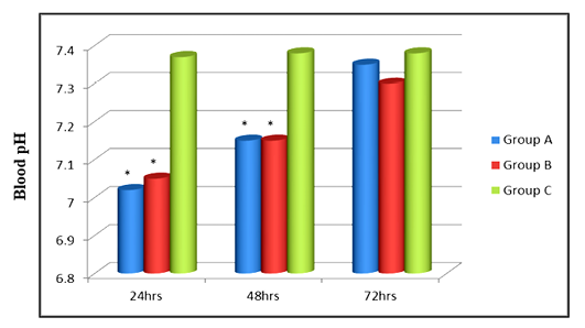
Figure 1: Effect of Cassia fistula and sodium bicarbonate+magnesium hydroxide on blood pH level in goats affected with lactic acidosis
*Significantly different at P < 0.05 as compare to control group C
Group A: Treated with Cassia fistula
Group B: Treated with sodium bicarbonate+magnesium hydroxide
Group C: Control group
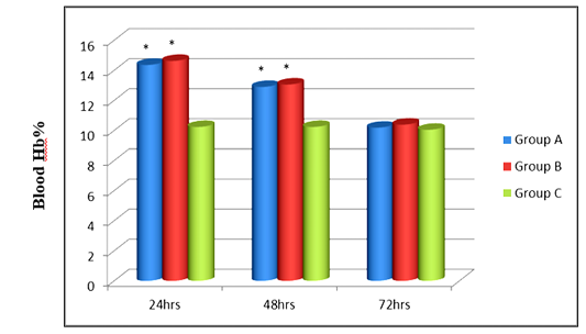
Figure 2: Effect of Cassia fistula and sodium bicarbonate+magnesium hydroxide on blood Hb (%) in goats affected with lactic acidosis
*significantly different at P < 0.05 as compare to control group C
Group A: Treated with Cassia fistula
Group B: Treated with sodium bicarbonate+magnesium hydroxide
Group C: Control group
-nificant (P < 0.05) difference during the 24hrs, 48hrs and 72hrs (Table 3). Goat in group B were treated orally with sodium bicarbonate+magnesium hydroxide as described before. Goat in this treatment group showed increased blood glucose level (157.14±36.49 mg/dL) returned to normal (91±8.67 mg/dL) within 72hrs. Total bilirubin, direct bilirubin, indirect bilirubin, ALT (SGPT) and alkaline phosphatase levels were increased. However, total bilirubin (0.77±0.04 mg/dL) and alkaline phosphatase were highly increased (439±3.65 U/L) at 24hrs, while minimum difference was observed in indirect bilirubin level (0.42±0.03). All the enzymes returned to normal with in 72hrs of treatment. Two way ANOVA revealed significant difference during the 24hrs, 48hrs and 72hrs at (P<0.05). On other
Table 3: Effect of Cassia fistula and sodium bicarbonate+magnesium hydroxide on blood glucose, total bilirubin, direct bilirubin, indirect bilirubin, ALT and alkaline phosphatase in goats affected with lactic acidosis
|
Group A (Treated with Cassia fistula) |
|||
| Variables |
24h** Mean ±SD |
48h** Mean ±SD |
72h** Mean ±SD |
| Glucose (mg/dL) |
132.87±37.91a |
99.87±11.63b |
81±5.60bc |
| Total bilirubin (mg/dL) |
0.79±0.03a |
0.72±0.03b |
0.60±0.04c |
| Direct bilirubin (mg/dL) |
0.29±0.03a |
0.27±0.02a |
0.20±0.02b |
| Indirect bilirubin (mg/dL) |
0.45±0.03a |
0.41±0.02b |
0.39±0.02b |
| ALT (SGPT) (U/L) |
38.5±2.56a |
35.25±3.28b |
30.62±1.92c |
| Alkaline phosphatase (U/L) |
438.87±14.74a |
411.25±12.90b |
326±17.82c |
| Group B ( Treated with sodium bicarbonate+magnesium hydroxide) | |||
| Glucose (mg/dL) |
157.14±36.49a |
131±35.32a |
91±8.67b |
| Total bilirubin (mg/dL) |
0.77±0.04a |
0.71±0.05b |
0.58±0.04c |
| Direct bilirubin (mg/dL) |
0.29±0.03a |
0.26±0.02b |
0.19±0.02c |
| Indirect bilirubin (mg/dL) |
0.42±0.03a |
0.40±0.02ab |
0.39±0.03bc |
| ALT (SGPT) (U/L) |
37.42±3.04a |
33±2.88b |
30.14±2.26bc |
| Alkaline Phosphatase (U/L) |
439±3.65a |
409.71±10.75b |
328.85±13.56c |
| Group C (Control) | |||
| Glucose (mg/dL) |
67.87±5.89a |
65.5±6.50a |
67±6.82a |
| Total bilirubin (mg/dL) |
0.56±0.03a |
0.57±0.02a |
0.56±0.02a |
| Direct bilirubin (mg/dL) |
0.19±0.01a |
0.19±0.01a |
0.19±0.01a |
| Indirect bilirubin (mg/dL) |
0.36±0.03a |
0.38±0.02a |
0.38±0.02a |
| ALT (SGPT) (U/L) |
27.87±2.41a |
28.62±2.44a |
28.12±2.16a |
| Alkaline Phosphatase (U/L) |
303.5±7.21a |
306.5±5.18a |
305.5±4.72a |
a-c Superscripts show significant difference
hand, goats in control group C goats did not show any significant change in glucose and liver function test results of total bilirubin, indirect bilirubin, direct bilirubin, ALT (SGPT) and alkaline phosphatase levels (Table 3).
Ruminal juice analysis of lactic acidosis goats treated by Cassia fistula and sodium bicarbonate+magnesium hydroxide
The ruminal juice examination of group “A” goats treated with Cassia fistula has shown change in the color, odor, and consistency from normal rapidly to yellowish, watery and soured. The observed decreased pH level (5.02±0.07) became normal (6.63±0.15) within the treatment of 72hrs. Two way ANOVA showed a highly significant (P < 0.05) difference during the 24hrs, and 72hrs (Figure 3). The ruminal juice analysis of group “B” goats showed that the pH level which was observed 5.02±0.07 returned to normal level 6.41±0.26 within the 72hrs of treatment. Two way ANOVA data analysis results showed significant (P < 0.05) difference during the 24hrs to 72hrs. The control group “C” goats did not show any change in ruminal juices and revealed all the parameters within normal range (Figure 3).
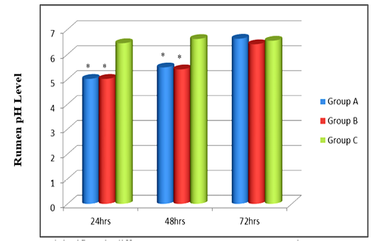
Figure 3: Effect of Cassia fistula and sodium bicarbonate+magnesium hydroxide on rumen pH in goats affected with lactic acidosis
*significantly different at P < 0.05 as compare to control group C
Group A: Treated with Cassia fistula
Group B: Treated with sodium bicarbonate+magnesium hydroxide
Group C: Control group
Efficacy of Cassia fistula and sodium bicarbonate+magnesium hydroxide against lactic acidosis in goats
Goats in group A were treated with Cassia fistula and in group B with sodium bicarbonate+magnesium hydroxide. These groups revealed the response of drugs i.e. 100% and 87.5% respectively (Figure 4).
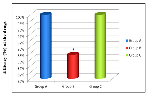
Figure 4: Efficacy (%) of Cassia fistula and sodium bicarbonate+magnesium hydroxide against lactic acidosis in goats
*significantly different at P < 0.05 as compare to control group C
Group A: Treated with Cassia fistula
Group B: Treated with Sodium bicarbonate+magnesium hydroxide
Group C: Control group
Discussion
In this study, goats were offered to take grains that induced lactic acidosis. All the goats were examined clinically and confirmed the lactic acidosis through the analysis of the ruminal fluid analysis and treated with Cassia fistula and sodium bicarbonate, magnesium hydroxide. All the goats had shown the all clinical signs of grain engorgements lactic acidosis similar to previously (Gonzalez et al., 2012). The decrease in body temperate, ruminal motility, pH and increase in heart rate, respiration rate and Hb% were observed as reported previously that goats ruminal bacteria suddenly causes fermentation to grain feed and produces the lactic acidosis (Hernández et al., 2014). The crushed wheat grains (50-60 gram) offered to sheep caused mortality. Therefore, all experimental goats had developed the lactic acidosis with in 12hrs and shown the typical clinical signs of lactic acidosis such as decrease in body temperature reported previously by (Ullah et al., 2012). However, lactic acidosis is also a leading to dehydration, ruminal motility ruminal pH and blood pH, these all signs were similar to reported previously and agreement with (Rodostitis et al., 2007).
Rapid fermentation of carbohydrate alters the ruminal function through proliferation of acid resistant bacteria and an increase in the production of volatile fatty acids and lactate, which cause a sharp drop in ruminal pH to (< 5.00) and dehydration were observed similar to previous study (Gozho et al., 2005; Gonzalez et al., 2010). The present experiment results were similar to the decreased in rumen pH that favors the growth of streptococci bovis and decrease in the gram negative bacteria and rumen protozoa, similar findings has been reported by several other researchers. The decrease in blood pH of lactic acidotic goats was found in close agreement with (Yang, 2012; Lascano et al., 2011).
In the present study of lactic acid affected goats, the predominant signs were observed anorexia, apathy, teeth grinding, ruminal stasis and increased respiration rate in group “A” and Group “B” with shallow and rapid breath in accordance to (Mohebbi et al., 2010). This increase in respiration rate was may be due to dehydration and stimulation of respiratory center by increased of carbon-dioxide (CO2) content in blood and decreased blood pH. While, increase in respiration rate and pulse rate was observed similar to reported previously (Haji et al., 2006). Goats had shown decreased blood circulating volume, heamoconcentration and severe dehydration in lactic acidosis, one goat died due to severe lactic acidosis, these signs were similar to the previous reports that increase in the release of histamine and absorption of the lipopolysaccharide endotoxins and other vasoactive agents attribute to rapid death in goats, these results are in agreements to the earlier reports that pH of (<5.00) is being lethal for goats (Chand et al., 2016).
All goats after the ingestion of grain had shown anorexia, dehydration, increase in ruminal fluid and decreased ruminal pH, these physiological changes lead to decrease in gram negative bacteria and increase in gram positive bacteria in the rumen resulting decrease in appetite, further these changes favor the lactobacilli to utilize more carbohydrates and to produce excessive amount of lactic acids and its isomers with lactate salt. These all caused increase in osmotic pressure significantly which result in the movement of excessive quantities of fluid in to the rumen and dehydration, these results are in close agreement with previously reports (Rodostitis et al., 2007).
Treatment of Lactic acidosis is always focused on the correction of ruminal and systemic acidosis and to inhibit the further production of lactic acid (Anderson and Rings, 2008). The clinical symptoms were returned to the normal after treatment with Cassia fistula (Amaltas) more rapidly than treated with Sodium bicarbonate, Magnesium hydroxide. All goats after the treatment with Cassia fistula (Amaltas) pass the feces within 3hrs, and started feeding within 12hrs of treatment, when pH began to increased, Cassia fistula (Amaltas) fruit seed pulp effect is considered to be the purgative action due to the presence of anthraquinone and mineral that increase the ruminal and intestinal motility, as anthraquinone is highly stimulating laxative that have direct effects on enterocytes and GIT smooth muscle that possibly induce a low-grade inflammation in the epithelium membrane of small and large intestine and stimulate the accumulation of water and electrolytes accumulation in the lumen of intestine, the feedback response starts in activation of prostaglandin-cyclic Adenosine monophosphate (cAMP) and Nitric oxide-cyclic (NO-cyclic), Guanosine monophosphate pathways (GMP pathways) and inhibition of Na+,K+-ATPase response (Agrawal et al., 2012).
Motility is a complex phenomenon affected both by local reflex (distension of GIT with food material or waste product) either through stretch receptors and the involvement of local hormones like cholecystokinin, ATP, kinins, Vasoactive protein, NO, intestinal peptides and PGs etc. In our present study fruit seed pulp of Cassia fistula (Amaltas) showed laxative action with an increase in intestinal fluid accumulation, intestinal motility and passing of the feces. These results were in similar to previously reported (Ding and Xu, 2006). In all goats with acidosis, ruminal fluid was observed with watery consistency and souring odor, these findings were in agreement with (Alam et al., 2014). The principles of the blood and ruminal pH reverted to the normal after the treatment with Sodium bicarbonate and Magnesium hydroxide more rapidly as compared to Cassia fistula treatment, the reason may be Dextrose 5% solution containing Sodium bicarbonate and Magnesium hydroxide orally was used for the correction of dehydration and stabilize the pH of rumen, these results suggested that using ruminal antacids orally to neutralize the ruminal acids and intravenous hypertonic Sodium bicarbonate are very effective to neutralize systemic acidosis and stable the animal to normal condition. After treatment with alkalizing agent sodium bicarbonate and magnesium hydroxide, lactic acid decreased more rapidly that stabilized ruminal pH between 6.2- 7.2, as reported before that normal return of rumen pH was the main factor for the early recovery to feed intake after the course of lactic acidosis (Rodostitis et al., 2007).
The blood glucose level was found increased after the ingestion of grains this indicated that grain feed was converted into highly fermentable action of the rumen microbial flora, these results are similar (Rahman, 2009). While liver function test like, total bilirubin, direct bilirubin, indirect bilirubin, ALT (SGPT) and alkaline phosphatase increased during the lactic acidosis in all goats, as reported before that In cattle being placed on a grain ration gradually on daily intake basis, hepatic cell damage and liver dysfunction occur even though dietary adaptation may have occurred in 2-3 weeks. The biochemical profile liver indicated that complete metabolic adaptation requires at least 40 days for adoption and decrease to damage to liver (Rodostitis et al., 2000). While increased level of ALT reflected hepatocellular damage which may be due to mild degeneration or necrosis to the hepatic cells, and increase in AST may be the reason of hepatocellular damage or by the degeneration of skeletal muscles, over-distention of rumen also impedes venous return to liver and heart, this impairs hepatic perfusion and further decrease in lactic acid utilization resulting development of systemic lactic acidosis (Arora et al., 2011). These all results are parallel to the study of Valmik et al. (2017) they reported that clinical signs after treatment in all acidotic goats disappeared as similar treatment was advocated by many earlier researchers (Chaudhary et al., 2009; Chakrabarti and Amalendu, 2006).
Conclusion
On the basis of present findings it could be concluded that the Cassia fistula administration in against lactic acidosis in remains most effective. It increases the ruminal and intestinal motility and rapidly restore all physiological parameters to normal range. It was further identified that Cassia fistula is very effective laxative to expel the ruminal ingesta containing grain and thus protect liver. It was also found that sodium bicarbonate+magnesium hydroxide were also helpful in restoring normal ruminal and blood pH but compared to Cassia fistula their efficacy is poor.
acknowledgements
Authors are thankful to the all staff members of Department of Veterinary Medicine, Sindh Agriculture University, Tandojam for providing research facility and conducive environment for current research project.
Conflict interests
The authors have declared that no competing interests exist.
authors contribution
Muhammad Qasim Koondhar: Performed research experiments, collected data, analyzed the data using statistical software, contributed in the conclusion, references and overall quality management of the manuscript.Asad Ali Khaskheli: Conceived the research idea, designed experiments, provided technical inputs at every step of study, wrote the research manuscript.
References


