Advances in Animal and Veterinary Sciences
Research Article
Advances in Animal and Veterinary Sciences. 1 (4S): 51 – 53Special Issue–4 (Progress in Research on Viruses and Viral Diseases)
Occurrence of Group a Rotavirus in Diarrhoeic Buffalo and Cow Calves, Madhya Pradesh, India
Aniruddha Udaykar1, Rakesh Sharda2*, Yashpal Singh Malik3, Varsha Sharma4, Nidhi Shrivastava5
- Wockhardt Limited, Aurangabad, Maharashtra, India
- Department of Microbiology, N.D.V.S.U., College of Veterinary Science and Animal Husbandry, Mhow, Madhya Pradesh, India
- Division of Biological Standardization, Indian Veterinary Research Institute, Izatnagar, India
- Department of Microbiology, College of Veterinary Science and Animal Husbandry, N.D.V.S.U., Jabalpur 482001, India
- Department of Pathology, College of Veterinary Science and Animal Husbandry, N.D.V.S.U., Madhya Pradesh, India
*Corresponding author:swaraksha@yahoo.com
ARTICLE CITATION:
Udaykar A, Sharda R, Malik YS, Sharma V, Shrivastava N. (2013). Occurrence of group a rotavirus in diarrhoeic buffalo and cow calves, Madhya Pradesh, India. Adv. Anim. Vet. Sci. 1 (4S): 51 – 53.
Received: 2013–11–17, Revised: 2013–12–17, Accepted: 2013–12–19
The electronic version of this article is the complete one and can be found online at
(
http://nexusacademicpublishers.com/table_contents_detail/4/162/html
)
which permits unrestricted use, distribution, and reproduction in any medium, provided the original work is properly cited
ABSTRACT
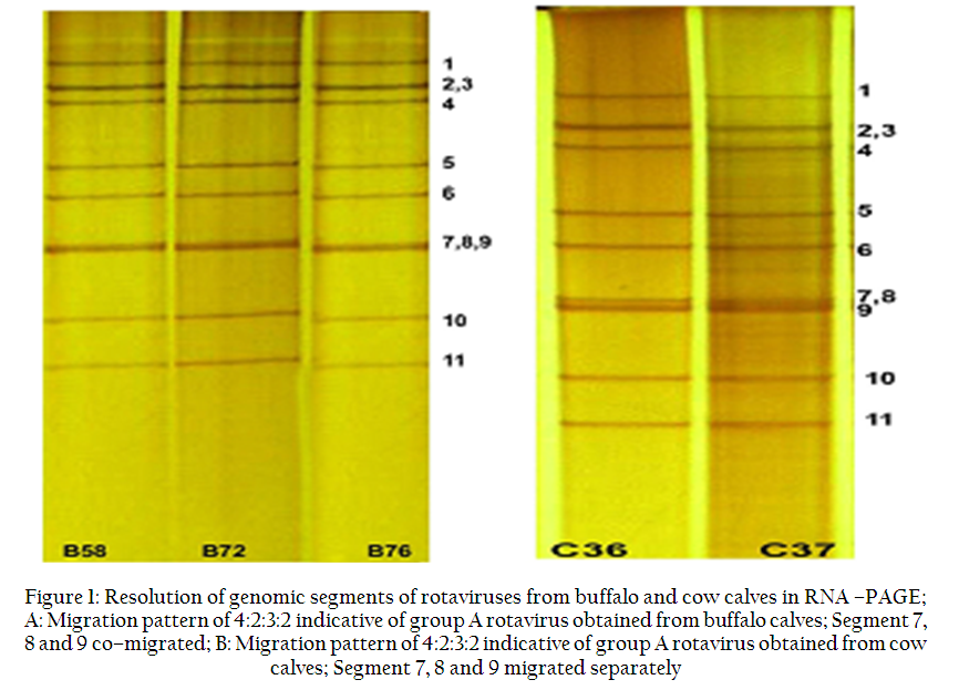
Virus induced gastrointestinal infections are amongst the most serious problems leading to huge economic losses particularly in developing countries. Amid gastro–enteric infections, rotavirus (RV) majorly dominates. We conducted a study to know the prevalence of RVs in cow and buffalo calves in the Malwa and Nimar agroclimatic zones of Madhya Pradesh, the central part of India. A total of 116 faecal samples collected from calves suffering with diarrhoea were subjected to electrophoretic and PCR based detection of RVs. The genomic migration pattern of viral RNA segments in 4:2:3:2 clusters and positive amplification of VP7 gene by RT–PCR confirmed the presence of RVs in 5 (4.3%) samples. Buffalo calves showed higher prevalence (4.76%) over cow calves (3.77%). These virus isolates were confirmed to be of group A with short electropherotype. The RVs recovered from buffalo calves showed different pattern of migration than cow calves in PAGE. The results confirmed the constant circulation of RV in dairy herds of this part of the country and warrants further studies to know about the circulating genotypes.
INTRODUCTION
Viral gastroenteritis is one of the most common diseases upsetting young animals internationally. It is one of the main causes of calf morbidity and mortality causing major economic loss in the dairy and beef herds. Gastroenteritis is multifactorial, involving both infectious and non–infectious causes. The known etiological agents include bacteria, viruses and protozoa. Amid all infectious causes, RVs are the principal etiological agent (Estes and Kapikian 2007; Dhama et al., 2009).
Rotaviruses comprise a genus within the family Reoviridae and the RV virion has a non–enveloped, complex, triple–layered capsid structure that surrounds 11 double–stranded RNA (dsRNA) genome segments. The laboratory diagnosis for RV is conventionally done by RNA–polyacrylamide gel electrophoresis (RNA–PAGE), electron microscopy, cultivation and isolation of virus in cell culture, antigen detection in faces by enzyme immunoassays (ELISA), latex agglutination test and molecular techniques involving RT–PCR (Minakshi et al., 2005). The present study records the prevalence of group A rotaviruses in buffalo and cow calves suffering with diarrhoea in Malwa and Nimar region of Madhya Pradesh, India.
MATERIALS AND METHODS
A total of 116 diarrhoeic faecal samples were aseptically collected from 63 buffalo and 53 cow calves reared in organized and unorganized farms of Malwa and Nimar regions of Madhya Pradesh, which comprise of eight revenue districts. The samples were collected in sterile test tubes containing PBS (pH7.2) and immediately transported on ice to laboratory. The faecal samples were re–suspended in 0.1M PBS, pH 7.2, to make 5% suspension, followed by centrifugation at 10,000 rpm for 30 min to remove coarse debris. Viral RNA extraction was done using guanidine isothiocyanate (GIT) lyses method as used in our previous study (Malik et al., 2012).
RNA–Polyacrylamide Gel Electrophoresis (RNA–PAGE)
Primary screening for presence of RV genome in samples was carried out by RNA–PAGE following the previously described procedure (Malik et al., 2012). Briefly, viral RNA extracted from faecal samples and suspended in 2x RNA sample buffer. It was electrophoresed at 100 volts in 12% resolving and 5% stacking gel; tris–glycine buffer 1X was used in electrophoresis. Silver staining of gel was done as described earlier (Savita et al., 2008).
Reverse Transcription–Polymerase Chain Reaction (RT–PCR)
All the samples detected positive in RNA–PAGE for RV genome were subjected to RT–PCR using primers used in our previous studies (Basera et al., 2010; Malik et al., 2011, Malik et al., 2012; Malik et al., 2013). Briefly, viral RNA extracted by GIT lyses method was subjected to RT–PCR using Bio–RT one step RT–PCR kit (Taurus Scientific, USA). The conditions for the RT–PCR were optimized using the reference bovine rotavirus strains. Finally using the standard conditions the PCR reactions were carried out in 0.2 ml thin walled PCR tubes in 20µl volumes 1µl (10pmol) forward primer, 1µl (10pmol) reverse primer, 12.5µl 2X RT–PCR Master Mix and 5.5µl of Nuclease free water (NFW). The PCR components were mixed and spin shortly. The RT–PCR conditions involved reverse–transcription at 48C for 60 min following an initial denaturation step at 94C for 5 min and 30 cyclic conditions of 1 min at 94C, 1 min at 42C, and 1 min at 72C). The PCR products were resolved by agarose gel electrophoresis using 1% agrose gel containing 0.5µg/ml ethidium bromide in 1× tris–acetate–EDTA (TAE) buffer in submarine electrophoresis apparatus (Biemetra, USA) at 12 V/cm. The reference RV samples were obtained from the Department of Microbiology, College of Veterinary Science and A.H. Jabalpur (M.P).
Isolation of E. coli
The faecal samples were also processed for isolation of E. coli, following the method of Edwards and Ewing (1972). The isolated strains were identified as E. colion the basis of biochemical and morphological characteristic as described by Barrow and Feltham (1993), and confirmed serologically by National Escherichia Tying Centre, CRI, Kasuli (H.P.).
RESULTS AND DISCUSSION
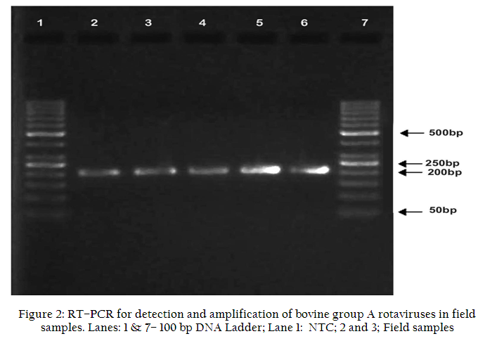
Out of 116 diarrhoeic fecal samples collected from cow and buffalo calves, RV was detected in 5 samples by RNA–PAGE (Figure 1 A, B) and confirmed by RT–PCR (Figure 2) yielding a prevalence rate of 4.3%.in Malwa region of Madhya Pradesh, India. Concurrent to our findings, Perez et al. (1998) also reported 7% prevalence rate of RV in diarrhoeic calves. On contrary, Kaminjolo and Adesiyun (1994) and Wani et al. (2007) recorded a higher prevalence rate of 27.7% and 18.7%, respectively. Rotavirus was detected in faecal samples of 3 buffalo and 2 cow calves out of 53 and 63 screened, respectively. Thus, a higher prevalence (4.76%) was recorded in buffalo than cow calves (3.77%), which is in corroboration with the reports from other regions of Madhya Pradesh (Sharma, 2004; Kusumakar et al., 2010). However, in northern India a higher prevalence was recorded in cow than buffalo calves (Jindal et al., 2000).
Samples were obtained from 36 male and 80 female calves. Sex–wise data analysis showed that prevalence was higher in female calves (5.0%) than males (2.77%), as also observed previously by Sharma (2004) in Madhya Pradesh during 2004–2006. Susceptibility was also evaluated for the different age groups of calves. Out of 21, 40, and 55 samples collected from 0–4, 4–8, and 8–12 week aged calves, respectively, RV was not detected in calves above two months of age. Only 3 and 2 samples from age group 0–4 and 4–8 week, respectively showed presence of virus. Minakshi et al. (2005) also reported that the susceptibility of bovine calves to RV decreases with age, probably due to loss of receptors on enterocytes.
In present study, RNA–PAGE was used as primary screening test to detect presence of RV in faecal samples. RNA–PAGE is a very sensitive and specific test for diagnosis of segmented genome viruses like RV and can detect about 108–1011 viral particles excreted per gram of faeces. The advantage lies in the fact that this technique can distinguish virus subgroup and variants and also detects non–cross reactive rotaviruses, which are missed in serological assays (Pereira et al., 1983). In this research pursuit electrophoretic migration pattern, characteristic of dsRNA genome of only group A rotavirus (4:2:3:2), was detected in all positive (5/116) faecal samples (Figure 1 A, B) collected from diarrhoeic cow and buffalo calves. Group A rotaviruses are one of the important causes of gastroenteritis in cow and buffalo calves worldwide including India causing measurable economical loss to the livestock industry (Janke et al., 1990; Kaminjolo and Adesiyun, 1994; Sunil–Chandra and Mahalingam, 1996; Jindal et al., 2000; Alfieri et al., 2004; Wani et al., 2007). Barman et al. (2004) reported emergence of a new genotype of bovine group B rotaviruses in India, but this subtype was not recorded in the present study.
Variations in RV genome, as depicted by different RNA migration patterns, can be studied through electropherotyping. Two distinct RNA segment migration patterns – "long”, defined by faster migration of 10th and 11th segments, and "short”, defined by slower migration of 10th and 11th segments of rotavirus RNA genome, have been identified (Espejo et al., 1977). In the present study all RV positive samples, from both cow (C36, C37) and buffalo (B58, B72, B76) calves, showed long electropherotype. The migration pattern of RNA segments in these samples was similar with minor variations. In all samples, segment 2 and 3 co–migrated or migrated very close. In buffalo samples, segment 7, 8 and 9 co–migrated, whereas in bovine samples segment 7 and 8 co–migrated and 9th segment migrated closely. Gulati et al. (1999), Jindal et al. (2000) and Wani et al. (2007) also reported variation in electropherotypes of bovine RV. Electropherotyping can be used as a detection method for RV infection and as a tool for studying its molecular epidemiology. Difference in migration profile of segment 7, 8 and 9 may be important as these segments encodes for the neutralizing antigens of RVs (Estes and Cohen, 1989). The faecal samples found positive for presence of RV by RNA–PAGE were further confirmed by RT–PCR in which VP7 gene was amplified. An expected amplicon of 208bp was obtained in these samples confirming the presence of RV.
The bacterial analysis of revealed isolation of sixty two (53.4%) strains of E. colifrom 116 fecal samples collected from cow and buffalo calves suffering with diarrhoea. Fifty six (90.32%) isolates were typed into 27 different ‘O’ serogroups, whereas 4 (6.45%) strains could not be typed and 2 (3.22%) were rough strains. Three of the faecal samples showed mixed infection of E. coliand RV. The E. colistrains belonged to serogroup O14, O22 and O138. Concurrent infection with RV and other enteropathogens is not uncommon (Perez et al., 1998; Cabalar et al, 2001). A combination of RV and E. colihas been reported to cause a more severe infection than either of the agents alone.
CONCLUSIONS
The present findings conclude that the occurrence of RV in buffalo and cow calves in central India is quite consistent. The study adds to the epidemiological data on RV in dairy herds of Madhya Pradesh, central India. However, still more elaborate studies should be conducted to estimate the gravity of the situation as well as the epidemiological understanding of contributing viral genotypes in future for the development and implementation of efficient immunization approaches, thereby controlling infection and reducing economic losses.
ACKNOWLEDGEMENTS
The authors are grateful to the Dean of the college for providing necessary facilities and to the Director, Central Research Institute, Kasauli, for serotyping E. coli.
REFERENCES
Alfieri AF, Alfieri AA, Barreieros MAB, Leite JPG and Richtzenhain LJ (2004). G and P genotype of group A rotavirus strains circulating in calves in Brazil, 1996–1999. Vet. Microbiol. 99: 167–173.
http://dx.doi.org/10.1016/j.vetmic.2003.10.029
PMid:15066719
Barman P, Ghosh S, Das S, Varghese V, Chaudhuri S, Sarkar S, Krishnan T, Bhattacharya SK, Chakrabarti A, Kobayashi N and Naik TN (2004). Sequencing and sequence analysis of VP7 and NSP5 genes reveal emergence of a new genotype of bovine group B rotaviruses in India. J. Clin. Microbiol. 42 (6): 2816–2818.
http://dx.doi.org/10.1128/JCM.42.6.2816-2818.2004
PMid:15184480 PMCid:PMC427839
Barrow GI and Feltham RKA (1993). Cowan and Steel's Manual for the Identification of Medical Bacteria. 3rd edn., Cambridge University Press, Cambridge. Pp. 140–43.
http://dx.doi.org/10.1017/CBO9780511527104
Basera SS, Singh R, Vaid N, Sharma K, Chakravarti S and Malik YPS (2010). Detection of Rotavirus Infection in Bovine Calves by RNA–PAGE and RT–PCR. Ind. J. Virol. 21(2):144–147.
http://dx.doi.org/10.1007/s13337-010-0017-9
PMid:23637494 PMCid:PMC3550708
Cabalar M, Boynukara B, Glhan T and Ekin IH (2001). Prevalence of rotavirus and Escherichia coli K95 and 'O'157: H7 in healthy dairy cattle herds in Van, Turkey. Turk – Veterinerlik – ve – Hay vancilik – Dergisi., 25 : 191–196.
Dhama K, Chauhan RS, Mahendran M and Malik SVS (2009). Rotavirus diarrhea in bovines and other domestic animals. Vet. Res. Comm. 33: 1–23.
http://dx.doi.org/10.1007/s11259-008-9070-x
PMid:18622713
Edwards R and Ewing WN (1972). Identification of Enterobacteriaceae. 3rd edn., Burgess Publishing Co., Minnesota. Pp. 211.
Espejo RT, Calderon E, Gonzalez N, Salomen A, Martuscelli A and Romero P (1977). Presence of two distinct types of rotavirus in infants and young children hospitalized with acute gastroenteritis in Mexico City. J. Infect. Dis. 139: 474–477.
http://dx.doi.org/10.1093/infdis/139.4.474
Estes M and Kapikian A (2007). Rotaviruses. In: Knipe DM, Howley PM, Griffin DE, Lamb RA, Martin MA, Roizman B, Straus SE, editors. Fields virology. Philadelphia: Kluwer Health/Lippincott, Williams and Wilkins. p. 1917–1974.
Estes MK and Cohen J (1989). Rotavirus gene structure and function. Microbiol. Rev. 53 (4): 410–449.
PMid:2556635 PMCid:PMC372748
Gulati BR, Nakagomi O, Koshimura Y, Nakagomi T and Pandey R (1999). Relative frequencies of g and p types among rotaviruses from Indian diarrheic cow and buffalo calves. J. Clin. Microbiol. 37(6): 2074–2076.
PMid:10325385 PMCid:PMC85038
Janke BH, Nelson JK, Benfield DA and Nelson EA (1990). Relative prevalence of typical and atypical strains among rotavirus from diarrheic pigs from convention herds. J. Vet. Diag. Invest. 2: 308–311.
http://dx.doi.org/10.1177/104063879000200410
PMid:1965637
Jindal SR, Maiti NK and Oberoi MS (2000). Genomic diversity and prevalence of rotavirus in cow and buffalo calves in northern India. Rev. Sci. Tech. 19: 871–876.
PMid:11107633
Kaminjolo JS and Adesinyun AA (1994). Rotavirus infection in calves, piglets, lambs and goat kids in Trinidad. Br. Vet. J. 150: 293–299.
http://dx.doi.org/10.1016/S0007-1935(05)80009-0
Kusumakar AL, Savita, Malik YPS, Minakshi and Prasad G (2010). Genomic diversity among group A rotaviruses from diarrheic children, piglets, buffalo and cow calves of Madhya Pradesh. Ind. J. Microbiol. 50(1): 83–88.
Malik YPS, Naveen Kumar, Kuldeep Sharma, AA Haq, Amit Kumar and Minakshi Prasad (2013). Sequence and phylogenetic analysis of bovine rotavirus isolates (G6 genotypes) from India. Adv. Vet. Anim. Sci. 1(1): 41–43.
Malik YPS, Sharma K, Vaid N, Chakravarti S, Chandrashekar KM, Basera SS, Singh R, Minakshi, Prasad S, Gulati BR, Bhilegaonkar KN and Pandey AB (2012). Frequency of group A rotavirus with mixed G and P genotypes in bovines: predominance of G3 genotype and its emergence in combination with G8/G10 types. J. Vet. Sci. 13(3): 271–278.
http://dx.doi.org/10.4142/jvs.2012.13.3.271
PMid:23006956 PMCid:PMC3467402
Malik YS, Chandrashekar KM, Sharma K, Haq AA, Vaid N, Chakravarti S, Batra M, Singh R and Pandey AB (2011). Picobirnavirus detection in bovine and buffalo calves from foothills of Himalaya and Central India. Trop. Anim. Health Product. 43(8): 1475–1478.
http://dx.doi.org/10.1007/s11250-011-9834-0
PMid:21479844
Minakshi, Prasad G, Malik YPS and Pandey R (2005). G and P genotyping of bovine group A rotaviruses in fecal samples of diarrheic calves by DIG–labelled probes. Indian J. Biotech. 4: 93–99.
Pereira HG, Linhares AC, Candeias JA and Glass RI (1983). National laboratory surveillance of viral agents of gastroenteritis in Brazil. Bull. Pan. Am. Health Organiz. 27: 224–233.
Perez E, Kummeling A, Janssen MMH, Jimenz C, Alvarado R, Caballero M, Donado P and Dwinger RH (1998). Infectious agents associated with diarrhoea calves in canton of Tilaran, Costa Rica. Prev. Vet. Med. 33: 195–205.
http://dx.doi.org/10.1016/S0167-5877(97)00038-X
Savita, Kusumakar AL, Malik YPS, Minakshi and Prasad G (2008). Detection and characterization of group A and D avian rotaviruses in India. Ind. J. Biotechnol. 7: 554–556.
Sharma R (2004). Isolation and molecular characterization of rotavirus associated with diarrhea in bovine calves. M.V.Sc & A.H. Thesis, JNKVV, Jabalpur.
Sunil–Chandra NP and Mahalingam S (1996). Isolation and subgrouping of rotaviruses from buffalo calves in Sri Lanka. Res. Vet. Sci. 60: 187.
http://dx.doi.org/10.1016/S0034-5288(96)90018-1
Wani SA, Bhat MA and Ishaq SM (2007). Molecular epidemiology of rotavirus in calves and lambs with diarrhoea in Kashmir Valley. Indian J. Virol. 18: 17–19.


