Advances in Animal and Veterinary Sciences
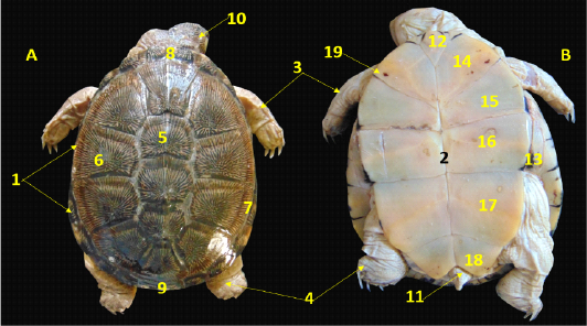
A. Dorsal view, B. Ventral view showing the external features of the female African helmeted turtle. 1. Carapase, 2.Plastron, 3.Forelimb, 4. Hind limb, 5.Vertebral scute, 6. Lateral costal scute, 7. Marginal scute, 8. Nuchal scute, 9. Supracaudal scute, 10.head, 11. Tail, 12. Gular Plastral scute, 13. Inguinal Plastral scute, 14. Humeral Plastral scute, 15. Pectoral Plastral scute, 16. Abdominal Plastral scute, 17. Femoral Plastral scute, 18. Anal Plastral scute, 19. Rathke’s pores
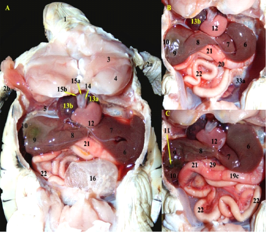
A; A photograph showing the ventral view of the coelomic cavity of African side neck turtle (fresh specimen) B; The urinary bladder was totally reflected, C; The right lobe of liver was slightly reflected.
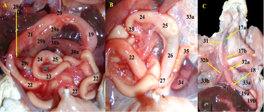
A; Photograph showing the pancreas parts after slight reflection of duodenum upward and rostrally, B; Magnified spleen in relation with the pancreas, C; The thoracic muscles were removed, neck was dissected and heart was removed.
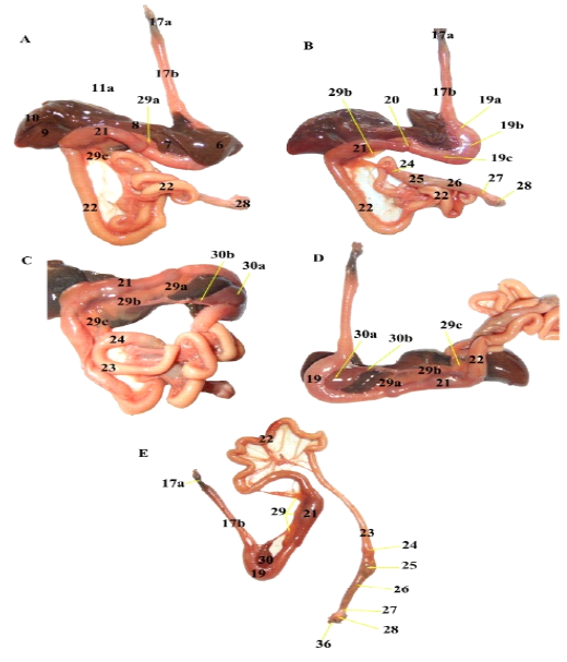
Photographs showing the Separated GIT; A; Ventral view, B; reflection of left lobe of liver, C; reflection of duodenum upward, D; parietal surface of stomach in relation to spleen, E; liver was removed.
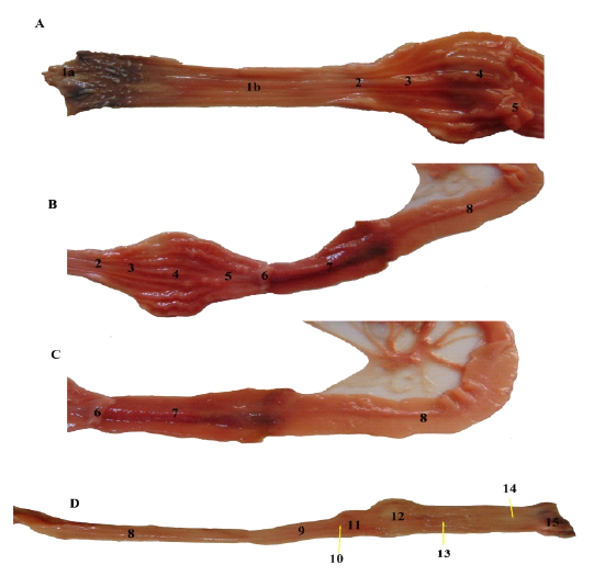
A, B, C &D: Photographs showing the opened gastrointestinal tract parts showing their mucosa.1a. 1st third of Oesophageal mucosa (pigmented and papillated with small pointed cones), 1b. Last two thirds of Oesophageal mucosa with numerous thin longitudinal folds, 2. Gastro oesophageal sphincter mucosa with small longitudinal folds without papillae. 3. Cardiac region of stomach with thin longitudinal mucosal folds, 4. Fundic part with thick corrugated longitudinal folds, 5. Pyloric region with few longitudinal folds. 6. Pyloric sphincter mucosa without any folds. 7. Duodenum smooth, dark red colored mucosa, 8. Jejunal mucosa, 9. Ileum mucosa, 10. Ileocecal valve mucosa, 11. Cecal smooth mucosa (saccular region), 12. Transverse colon smooth mucosa, 13. Descending colon mucosa with rectilinear folds, 14. Rectal smooth mucosa, 15. Cloacal mucosa.
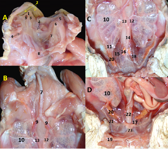
A. Roof and floor of oropharynx; B, C, D; anterior, middle, posterior parts of coelomic cavity of female African side neck turtle, respectively. 1. Hard palate, 2. Villiform papillae, 3. Internal Choanae, 4. Opening of Eustachian tube, 5. Tongue, 6. Glottis.7. Trachea, 8. Cut oesophagus, 9. Extra pulmonary bronchus, 10. Lung, 11. posterior angle of lung, 12. Left aorta 13. Right aorta, 14. Dorsal aorta 15. Kidney, 16. Adrenal gland, 17. Ureter, 18. Segmental artery, 19. Urinary bladder (reflected), 20. Ovary, 21. Oviduct, 22. Descending Colon, 23. Cloaca.
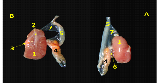
Photographs showing kidney in relation with ovary, A. Dorsal view; B. Ventral view, 1. Kidney, 2. Segmental artery, 3. Adrenal gland, 4. Ovary, 5. Oviduct, 6. Mesovarium, 7. Mesosalpinx.


