Advances in Animal and Veterinary Sciences
Research Article
Advances in Animal and Veterinary Sciences. 1 (3S): 12 – 16Special Issue–3 (Epidemiology and Animal Disease Investigations)
Detection of Bovine Herpesvirus–l (BHV–l) Infection in Cattle by Antigen Detection ELISA and Multiplex PCR
Rashmi Singh1*, Amit Kumar Verma1, Barkha Sharma1, Sharad Kumar Yadav2
- Department of Veterinary Epidemiology and Preventive Medicine, Uttar Pradesh Pandit Deen Dayal Upadhayay Pashu Chikitsa Vigyan Vishwavidyalaya Evum Go–Anusandhan Sansthan (DUVASU), Mathura, India – 281001
- Department of Veterinary microbiology and Immunology, Uttar Pradesh Pandit Deen Dayal Upadhayay Pashu Chikitsa Vigyan Vishwavidyalaya Evum Go–Anusandhan Sansthan (DUVASU), Mathura, India – 281001
*Corresponding author:rashmifauzdar@gmail.com
ARTICLE CITATION:
Singh R, Verma AK, Sharma B, Yadav SK (2013). Detection of bovine herpesvirus–l (BHV–l) infection in cattle by antigen detection ELISA and multiplex PCR. Adv. Anim. Vet. Sci. 1 (3S): 12 – 16.
Received: 2013–11–13, Revised: 2013–12–16, Accepted: 2013–12–17
The electronic version of this article is the complete one and can be found online at
(
http://nexusacademicpublishers.com/table_contents_detail/4/158/html
)
which permits unrestricted use, distribution, and reproduction in any medium, provided the original work is properly cited
ABSTRACT
In present cross sectional study, nasal swabs were collected from cattle of organized and unorganized herds (n=333) from seven districts of Uttar Pradesh state, and examined for presence of BHV–1 using antigen detection by ELISA and subsequently confirmed by multiplex–Polymerase chain reaction (multiplex–PCR). Overall percent positivity of BHV–1 antigen in cattle of Uttar Pradesh examined was 11.1% (37/333). The percent positivity of BHV–1 was higher in organized herd (15.38%) than that of unorganized herd (4.4%). All the 37 samples positive by ELISA were processed further for molecular characterization using gB and gC based multiplex PCR. Out of 37 samples, 20 (54.0%) samples were positive with gB and gC based multiplex PCR. Among these, 20 positive samples by PCR, 11(29.7%) samples were positive with both gB and gC gene primers of multiplex PCR, while 6 (16.2%) samples were positive only with gB gene primer and 3 (8.1%) samples were positive only with gC gene primer. From the study, it can be concluded that the BHV–1 virus is circulation in cattle of Uttar Pradesh, India, which causes significant economic impact to dairy industry and export. Although this study was performed using less number of sample from limited geographical area, so a detailed study should be performed using more number of samples from vast geographical area.
INTRODUCTION
Worldwide, livestock health problems particularly of reproductive system leads to direct and indirect economic losses to dairy industry by abortions, still births, early embryonic mortality, retention of placenta, poor fertility, and loss of production (Poulsen and McGuirk, 2009; Gay and Barnouin, 2009; Raaperi et al., 2012). Among these health problems majority are of infectious origin such as brucellosis, leptospirosis, campylobacteriosis, listeriosis and infectious bovine rhinotrachitis (Kumar et al., 2009; Verma et al., 2014). Infectious bovine rhinotracheitis (IBR) is a major, economically important and emerging disease of cattle, caused by bovine herpesvirus–1 (BHV–1), causing various clinical syndromes viz., respiratory, reproductive (vulvovaginitis or balanoposthitis), conjunctivitis, encephalitis and generalized systemic infections (Gibb and Rweyemamu, 1977; Straub, 1991; Nandi et al., 2009; Jacevicius et al., 2010; Verma et al., 2014).
BHV–1 genome consist of linear, double stranded DNA of about 1,36,000 base pairs, enveloped with glycoproteins spikes on its surface and its structure is typical of herpesviruses of group D (Roizman, 1992). There are 8 known glycoproteins viz., gB, gC, gD, gF, gH, gI, gK and gL. Out of these, gC, gD, gE, gG, gI, UL49h and thymidine kinase genes are involved in viral virulence (Smith, 1991; Smith et al., 1994; van Engelenburg et al., 1994; Kaasheek et al., 1998). Studies show that the BHV–1 glycoproteins gB, gC, gD, gE, gH, gk, gL are required for virus entry (Schroder and Keil 1999). BHV–1 isolates were classified into subtype 1 (BoHV–1.1) and BoHV–1.2 according to distinct restriction enzyme profiles of the genomes. In India, disease was first time reported in Uttar Pradesh state and since then many reports have been published regarding its occurrence in different states of the country (Mehrotra et al., 1976; Renukaradhya, 1996; Rajkhowa et al., 2004; Sunder et al., 2005; Ganguly et al., 2008; Nandi et al., 2010; Verma et al., 2014). For effective control of disease, early and confirmatory diagnosis is very important. Recently, emphasis has been given to reduce the time required for diagnosis of infections. Virus isolation in cell culture is most frequently used for diagnosing BHV–1 but it is laborious, time consuming, and requires samples of good quality. Hence alternative techniques like ELISA and polymerase chain reaction have been tried. The present manuscript describes the epidemiological studies of BHV–1 using antigen based ELISA and multiplex PCR in cattle of Uttar Pradesh, India.
MATERIALS AND METHODS
Study Design, Area and Sample Collection
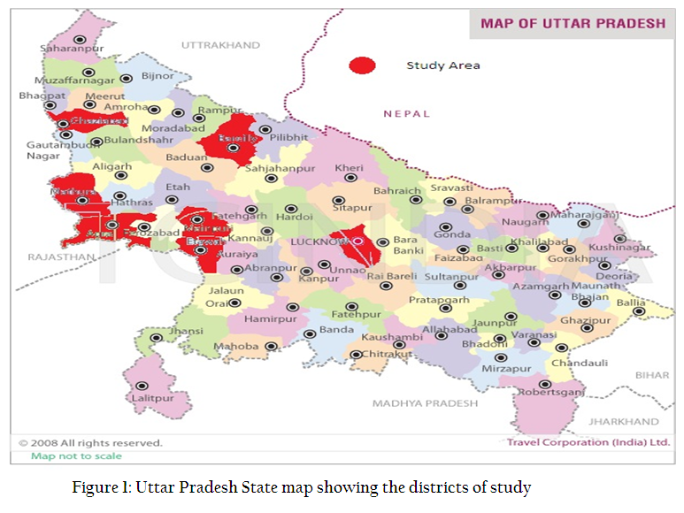
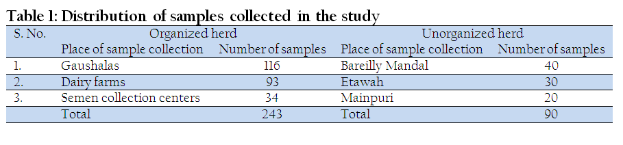
This cross–sectional study was conducted in seven districts (Agra, Bareilly, Etawah, Ghaziabad, Lucknow, Mainpuri and Mathura) of Uttar Pradesh, India (Figure 1). A total of 333 nasal swabs were collected from cattle of 1–4 years of age. Samples were taken from different farms, semen collection centers and gaushalas in Uttar Pradesh. Among these 333 nasal swabs, 243 samples were taken from organized herd and 90 samples were taken from unorganized herd (Table 1). The nasal swabs were dipped in Eagle’s MEM containing antibiotics, thoroughly shaked and centrifuged at 1000g for 10 min at 4oC. The supernatants from nasal swabs were taken for ELISA and viral DNA extraction.
Laboratory Examination
The laboratory analysis was conducted at Department of Veterinary Epidemiology and Preventive Medicine, Uttar Pradesh Pandit Deen Dayal Upadhayay Pashu Chikitsa Vigyan Vishvidhyalaya Evum Go–Anusandhan Sansthan (DUVASU), Mathura, India by antigen detection (using sandwich ELISA) and viral DNA detection (using Multiplex polymerase chain reaction).
Antigen Detection by Sandwich–ELISA
The nasal samples were tested to detect presence of BHV–1 antigen, using a commercially available sandwich enzyme linked immunosorbent assay (ELISA) kit (BIO–X Pulmotest BHV–1 ELISA kit) following manufacturer’s recommendations.
Viral DNA detection by Multiplex PCR
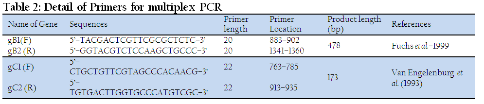
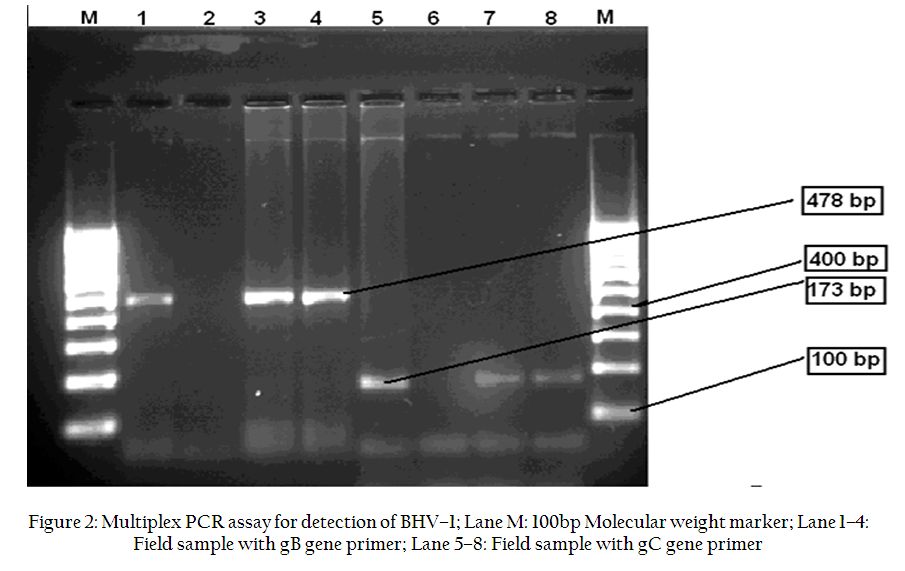
The samples, which were positive by antigen detection sandwich ELISA, were processed further for Multiplex PCR using the specific primers (Table 2) For amplification in thermocycler (Techne, Japan) an initial denaturation (5 min at 95oC) was followed by 35 cycles of denaturation (1 min at 94oC), annealing (1 min at 60oC) and extension (1 min at 72oC) and a step of final extension for 10 minutes at 72oC. The amplicons of 173bp and 478bp were visualized under UV illuminator after agarose gel electrophoresis (5 volts/cm) using 1.5% agarose made in 0.5X Tris–borate buffer (TBE) containing ethidium bromide (0.5µg ml–1).
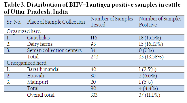
In the present cross–sectional study, a total of 333 nasal samples (243 samples from organized herd and 90 samples from unorganized herd) of cattle from seven districts of Uttar Pradesh state, India were analyzed for presence of BHV–1 antigen. Overall positive of BHV–1 antigen in cattle of Uttar Pradesh examined was 11.1% (37/333). In samples from organized herd screened, 15.38% (33/243) exhibited positive reaction, while 4.4% (4/90) nasal swab samples from unorganized herds exhibited positive reaction (Table 3).
With variability in the percentage presence of IBR antigen (virus) in various secretions of the animals having the clinical history of the disease. In the present study, higher percent positivity of BHV–1 was observed in organized herd in comparison to that of unorganized herd. This might be due to spread of infection from one animal to other either by close contact between the animals or during natural service with infected bulls as well as poor hygiene practices like improper disposal of aborted fetuses, foetal membranes, uterine and vaginal discharges.
All the 37 samples, positive by ELISA, were processed further for molecular characterization using gB and gC based multiplex PCR. Out of 37 samples, 20 (54.0%) samples were positive with gB and gC based multiplex PCR. Among these, 20 positive samples by PCR, 11(29.7%) samples were positive with both gB and gC gene primers of multiplex PCR, while 6 (16.2%) samples were positive only with gB gene primer and 3 (8.1%) samples were positive only with gC gene primer (Table 4; Figure 2).
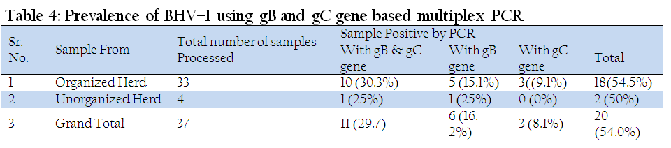
Similarly, various researchers used PCR for molecular detection of BHV–1 in nasal swabs with results range from 25% to 66.6% (Vilcek et al., 1995; Gee et al., 1996; Rola et al., 2005). However in the present study, a multiplex PCR was developed for detection of gB and gC genes. The already known primers of these genes were optimized in the multiplex reactions to perform the multiplex PCR. Results showed percent positivity of gB and gC positive samples was 29.7%, while percent positivity of only gB and only gC gene was 16.2% and 8.1%, respectively. Very few multiplex PCR were developed incorporating these two genes based primers. However the gC gene based primer can detect the virus in latency (Winkler et al., 2000).
CONCLUSION
From the study, it can be concluded that the BHV–1 virus is circulation in cattle of Uttar Pradesh, India, which causes significant economic impact to dairy industry and export. Although this study was performed using less number of sample from limited geographical area, so a detailed study should be performed using more number of samples from vast geographical area.
ACKNOWLEDGEMENT
This work was supported by Indian Council of Agriculture Research (ICAR) under Niche Area of Excellence project. The authors are highly thankful to Head, Department of Veterinary Microbiology and Immunology; Department of Veterinary Epidemiology and Preventive Medicine, Dean, College of Veterinary Sciences and Animal Husbandry, and Hon’ble Vice chancellor, Uttar Pradesh Pandit Deen Dayal Upadhayay Pashu Chikitsa Vigyan Vishvidhyalaya Evum Go–Anusandhan Sansthan (DUVASU), Mathura, India; for providing all the necessary support and facilities for conducting this study. The authors are highly thankful to animal owners, who allowed their animals for participation in this study.
REFERENCES
Ackermann M and Muller HK (1990). Eradication of infectious bovine rhinotracheitis in Switzerland: review and prospects. Vet. Microbial. 23: 365–370.
http://dx.doi.org/10.1016/0378-1135(90)90168-U
Adler Storthz, K, Kendall, Kennedy CRC, Henkel RD,and Dreesman GR(1983). Biotin–avidin–amplified enzyme immunoassay for detection of herpes simplex virus antigen in clinical specimens. J. Clin Microbiol.18:1329–1334.
PMid:6317711 PMCid:PMC272902
Ganguly, S, Mukhopadhayay, SK and Paul, I (2008). Studies on seroprevalence of Infectious Bovine Rhinotracheitis in cattle population of West Bengal. Indian J. Comp. Microbiol. Immunol. Infect. Dis. 29(1&2): 12–16
Gay, E, Barnouin, J (2009). A nation–wide epidemiological study of acute bovine respiratory disease in France. Preventive Veterinary Medicine 89: 265–271.
http://dx.doi.org/10.1016/j.prevetmed.2009.02.013
PMid:19297044
Gee, A L W De, Wagter, LHA and Hage, JJ (1996). The use of a polymerase chain reaction assay for the detection of bovine herpesvirus 1 in semen during a natural outbreak of infectious bovine rhinotracheitis. Vet. Microbiol. 53: 163–168.
http://dx.doi.org/10.1016/S0378-1135(96)01244-8
Gibbs EPJ and Rweyemuma MM (1977). Bovine herpesviruses. Part1. Bovine herpes virus 1. Vet Bull 47: 317–343.
Fuchs M, Hubert P, Detterer J, and Rziha HJ(1999). Detection of Bovine Herpesvirus Type 1 in Blood from Naturally Infected Cattle by Using a Sensitive PCR That Discriminates between Wild–Type Virus and Virus Lacking Glycoprotein E. Journal of clinical microbiology, 37, (8): 2498–2507.
PMid:10405392 PMCid:PMC85268
Jacevicius, E, Salomskas, A, Milius, J, Petkevicius, S, Jaceviciene, I, Pridotkas, G, Mockeliunas, R, Malakauskas, A and Morkunas, M (2010). Five year serological study of Bovine Herpesvirus type–1 in cattle in Lithuania. Bull Vet Inst Pulawy 54: 289–292.
Kaashoek, MJ, Rijsewijk, FA, Ruuls, RC, Keil, GM, Thiry, E, Pastoret, PP, Van Oirschot, JT (1998). Virulence, immunogenicity and reactivation of bovine herpesvirus 1 mutants with a deletion in the gC, gG, gI, gE, or in both the gI and gE gene. Vaccine 16: 802–801.
http://dx.doi.org/10.1016/S0264-410X(97)00269-7
Kumar, N, Pal, BC, Yadav, SK, Verma, AK, Jain, U and Yadav, G (2009). Prevalence of Bovine Brucellosis in Uttar Pradesh, India. Journal of Veterinary Public Health, 7(2): 129–131.
Kupferschmied HU and Kihm U (1986). Transmission of IBR/IPV virus in bovine semen: a case report. Theriogenology 25: 439–443.
http://dx.doi.org/10.1016/0093-691X(86)90052-X
Land, SA, Skurrie IJ, and GL Gilbert GL(1984). Rapid diagosis of herpes simplex virus infections by enzyme linked immunosorbent assay. J. clin. Microbiol. 19:865–869.
PMid:6088571 PMCid:PMC271200
Mehrotra, ML, Rajya, BS and Kumar, S (1976). Infectious bovine rhinotracheitis (IBR) – keratoconjunctivitis in calves. Indian Journal of Veterinary Pathology 1: 70–73.
Miller JM (1991). The effect of IBR virus infection on reproductive function of cattle. Vet Med 86: 95–98.
Morgan, MA, and Smith, TF (1984). Evaluation of an enzyme –linked immunosorbent assay for the detection of herpes simplex virus antigen. J. Clin. Microbiol. 19:730–732.
PMid:6088568 PMCid:PMC271173
Nandi S, Kumar M, Manohar M and Chauhan RS (2009). Bovine herpes virus infections in cattle. Anim Health Res Rev. 10(1):85–98.
http://dx.doi.org/10.1017/S1466252309990028
PMid:19558751
Nandi S, Kumar M, Yadav V and Chander V (2010). Serological Evidences of Bovine Herpesvirus–1 Infection in Bovines of organized Farms in India. Transbound Emerg Dis. doi: 10.1111/j.1865–1682.2010.01185.x.
http://dx.doi.org/10.1111/j.1865-1682.2010.01185.x
Poulsen, KP, McGuirk, SM, (2009). Respiratory disease of the bovine neonate. The Veterinary Clinics of North America. Food Animal Practice 25: 121–137.
http://dx.doi.org/10.1016/j.cvfa.2008.10.007
PMid:19174286
Raaperi, K, Bougeard, S, Aleksejev, A, Orro, T and Viltrop, A (2012). Association of herd BHV–1 seroprevalence with respiratory disease in young stock in Estonian dairy cattle. Research in Veterinary Science 93: 641–648.
http://dx.doi.org/10.1016/j.rvsc.2011.10.015
PMid:22100246
Rajkhowa, S, Rajkhowa, C, Rahman, H and Bujarbaruah, KM (2004). Seroprevalence of infectious bovine rhinotracheitis in mithun (Bos frontalis) in India. Review Scientifiic Tech Office Intertnational Epizooties 23: 821–29.
Renukaradhya, GJ, Rajasekhar, M and Raghavan, R (1996). Prevalence of infectious bovine rhinotracheitis in Southern India. Rev. Sci. Tech. Off. Int. Epiz. 15: 1021–1028.
Roizman B (1992). The family Herpesviridae:an update.Arch Virol. 123: 425–449.
http://dx.doi.org/10.1007/BF01317276
Rola, J, Larska, M and Polak, MP (2005). Detection of bovine herpesvirus 1 from an outbreak of infectious bovine rhinotracheitis. Bulletin of the Veterinary Institute in Puawy. 49:267–271.
Schroder, C and Keil, GM (1999). Bovine herpesvirus 1 requires glycoprotein H for infectivity and direct spreading and glycoproteins gH (W450) and gB for glycoprotein D–independent cell–to–cell spread. Journal of General Virology 80: 57–61.
PMid:9934684
Singh, A and Sinha, BK (2006). Seroprevalence of infectious bovine rhinotracheitis (IBR) in cattle in Bihar. Indian J. Comp. Microbiol. Immunol. Infect. Dis. 27(2): 107–108.
Smith, GA (1991). Analysis of BHV1 TK genes and construction of an attenuated vaccine. PhD University of Queensland.
Smith, GA, Young, PL, Rodwell, BJ, Kelly, MA, Storie, GJ, Farrah, CA, Mattick, JS (1994). Development and trial of a bovine herpesvirus 1–thymidine kinase deletion virus as a vaccine. Australian Veterinary Journal 71: 65–70.
http://dx.doi.org/10.1111/j.1751-0813.1994.tb03329.x
PMid:8198509
Straub OC (1991). BHV–1 infections: relevance and spread in Europe. Comp Immunol Microbiol Infects Dis 14: 175–186.
http://dx.doi.org/10.1016/0147-9571(91)90130-6
Sunder, J, Rai, RB, Kundu, A, Chatterjee, RN, Senani, S and Jeyakumar, S (2005). Incidence and prevalence of livestock diseases of Andaman and Nicobar islands. Indian Journal of Animal Sciences. 75 (9): 1041–1043.
Van Engelenburg, FA, Kaashoek, MJ, Rijsewijk, FA, Van den, B.L, Moeman, A,Gielkens, AL, Van Oirschot, JT (1994). A glycoprotein E deletion mutant of bovine herpes virus 1 is avirulent in calves. Journal of general virology 75:2311–2311118.
http://dx.doi.org/10.1099/0022-1317-75-9-2311
PMid:8077929
Van Engelenburg, FAC, Maes, RK, Van Oirschot, JT, and Rijwijk, FAM (1993). Development of a rapid and sensitive polymerase chain reaction for the detection of bovine herpes virus type–1in bovine semen. Journal of clinical Microbiology. 31: 3129–3135.
PMid:8308103 PMCid:PMC266363
Verma, AK, Kumar, A, Sahzad, Reddy, NCR and Shende, AN (2014). Sero–prevalence of Infectious Bovine Rhinotracheitis in dairy animals with reproductive disorders in Uttar Pradesh, India. Pak. J. Biol. Sci. DOI 10.3923/pjbs/2014.
Vilcek, S, Nettleton, P F and Herring, AJ (1995). Detection of bovine herpesvirus1in clinical samples by the polymerase chain reaction. Dtsch. Tierarztl. Wochenschr. 102: 249–250.
PMid:8582261
Winkler MTC, Doster A and Jones C (2000). Persistent and reactivation of Bovine herpes virus1 in tonsils of latently infected Calves. Journal of virology. 74 (11): 5337–5346
http://dx.doi.org/10.1128/JVI.74.11.5337-5346.2000
PMid:10799611 PMCid:PMC110889
Yolken, RH 1982. Enzyme immunoassays for the detection of infectious antigens in body fluids: current limitations and future prospects. Rev. Infect. Dis. 4: 35–68.
http://dx.doi.org/10.1093/clinids/4.1.35
PMid:6803327


