Advances in Animal and Veterinary Sciences
Short Communication

The Pathogenic Effect of Eimeria on Rabbits of the Soviet Chinchilla Breed and its Hybrids with the Californian Breed
Karina Sidorenko1*, Manya Mkrtchyan1, Yuri Kuznetsov1, Ekaterina Klimova2
1Saint Petersburg State University of Veterinary Medicine, Saint Petersburg, Russia; 2Izhevsk State Agricultural Academy, Izhevsk, Russia.
Abstract | Questions of the influence of associations of protozoa of the genus Eimeria, depending on the intensity of infection on the organism of rabbits of various breeds, remain relevant. The object of the study was rabbits of the Soviet chinchilla breed (SCh * SCh) and its hybrid with the Californian breed (SCh * C) at the age of 40 days. As part of the experiment, formed four groups of analogs of rabbits, six animals in each: purebred (SCh * SCh) and hybrids (SCh * C) in the experimental (invasive) and control (intact) groups. The rabbits of experimental groups infected by a dose of 50-60 thousand oocysts per head with the associations of the Eimeria species E. Perforans and E. irresidua. To establish the pathogenic effect of parasites, we determined the meat productivity, carried out a veterinary and sanitary assessment of carcasses and linear measurements of the length of the intestines, as well as histological studies of the walls of the small and large intestines, liver and spleen of rabbits of these breeds. The research results showed that with a low infection rate of 50-60 thousand oocysts per head, the maximum live weight gain and slaughter yield of carcasses decrease in rabbits of a hybrid breed (SCh x C), despite the preservation of the marketable appearance of the body. In histological sections of the small intestine, at the sites of merontslocalization and merozoites, has found damaged enterocytes, and accumulations of lymphocytes, and eosinophils.
Keywords | Rabbits, Eimeria, Breed, Soviet chinchilla, Californian, Hybrid
Received | July 17, 2020; Accepted | August 28, 2020; Published | September 20, 2020
*Correspondence | Karina Sidorenko, Saint Petersburg State University of Veterinary Medicine, Saint Petersburg, Russia; Email: doooctor@yandex.ru
Citation | Sidorenko K, Mkrtchyan M, Kuznetsov Y, Klimova E (2020). The pathogenic effect of eimeria on rabbits of the soviet chinchilla breed and its hybrids with the Californian breed. Adv. Anim. Vet. Sci. 8(s2): 7-11.
DOI | http://dx.doi.org/10.17582/journal.aavs/2020/8.s2.7.11
ISSN (Online) | 2307-8316; ISSN (Print) | 2309-3331
Copyright © 2020 Sidorenko et al. This is an open access article distributed under the Creative Commons Attribution License, which permits unrestricted use, distribution, and reproduction in any medium, provided the original work is properly cited.
INTRODUCTION
The domestic rabbit (Oryctolagus cuniculus domesticus L.) is a small animal belonging to the order Lagomorphs. Based on structural features of the digestive system, it is classified as a pseudo-ruminant capable using a large amount of roughage, having a sufficiently developed cecum, as part of large intestines, for efficient microbial digestion (Ayan et al., 2020; Hamid et al., 2019).
The rabbit easily raised to produce high-quality meat known to be healthier than many other farm animals. Since it contains many polyunsaturated fatty acids and essential amino acids, it is an excellent source of animal protein. Rabbit meat also provides high calorie content but low levels of low-density lipoprotein and cholesterol, which is attracting more and more healthy consumers.
Rabbit breeding is very promising since the rabbit is highly productive. Based on the number of offspring obtained, short gestation and lactation periods, these animals have high fertility. Also, rabbits are easy to transport and do not require a large amount of feed and the rebuilding of large livestock complexes. They provide meat products, fur, and leather (Hamid et al., 2019).
That is why global production and consumption of rabbit products is are inevitably increasing from 1.22 million tons in 2010 to almost 1.43 million tons in 2016 (Hamid et al., 2019; Petrova, 2019).
Today, in 70 countries of the world, more than 305 breeds and species of rabbits are bred, which differ in their economic and purposeful purposes. In Eurasian countries, these animals, used mainly to obtain high-quality dietary meat (Bharathy et al., 2014).
The most significant production of rabbit meat in Asia has established in China, which provides 80% of Asia’s needs. About 180 million rabbits raised in Europe. Rabbit meat is the sixth most consumed meat after broilers, laying hens, trout, salmon, and pigs (Hamid et al., 2019).
In addition to a source of meat, rabbits, as, for example, in the United States, can be used to obtain fur raw materials, as a laboratory or decorative pets (Bharathy et al., 2014; Petrova, 2019).
Aymeriosis, as one of the most common parasitic diseases of the digestive system in rabbits, causes significant harm to productive livestock (Fagundes et al., 2005; Maziz-Bettahar et al., 2018).
The most critical indicators of meat productivity are slaughter weight, which characterizes the mass of an animal’s carcass without the head, legs, skin, and internal organs (except for the kidneys), and slaughter yield - the percentage of slaughter weight to the weight of the animal before slaughter.
Due to the rather rapid development of these pathogens, a short life cycle, the absence of an intermediate host, high reproductive properties, the greatest danger of eimeria presented to young farm animals, for which the disease often ends in death (Bezrukova et al., 2016; Lutfullin et al., 2016).
Protozoa directly affect the production potential of infected rabbits due to high mortality, stunted growth, and reduced feed conversion rates, resulting in high economic losses to the industry each year. Besides, in a subclinical form, they can lead to a decrease in the immune status of animals, which opens the way to secondary infections (Yin et al., 2016).
Aymeriosis is one of the main items of losses due to lost weight gain, livestock mortality, a high tendency to infection of all existing livestock in a short period, as well as high costs for long-term and often ineffective antiparasitic measures (Yatusevich, 2012).
According to many researchers, eimeria are always present on rabbit farms, are ubiquitous in the environment,and are practically indestructible (Hamid et al., 2019). The high viability of oocysts, as well as their ability to survive after contact with disinfectants in concentrations and exposures that destroy pathogenic microorganisms, is a severe problem in the fight against invasive animal diseases (Shkromada et al., 2019).
In protozoal diseases of the gastrointestinal tract, general clinical signs are and manifested by decreased appetite, depression, weight loss, diarrhea, and anemia of the mucous membranes. Feces may contain blood or mucus. However, any of these traits may be absent in adult rabbits, which are mainly parasitic carriers (Shkromada et al., 2019).
The causative agents of this invasive disease, most of which localized into the gastrointestinal tract, affect the cells of the limb epithelium, sometimes involving the intestinal lining of the intestinal mucosa in the process. That leads to disruption of digestion and absorption processes and accompanied by a decrease in both meat productivity and the quality of fur raw materials. Erosion and ulceration appear in the epithelial lamina of the intestinal mucosa leading to poor absorption of nutrients, electrolyte imbalance, anemia, and dehydration (Bezrukova et al., 2016; Mkrtchyan et al., 2019).
In our studies, we set out to carry out a comparative analysis of the pathogenic effect of protozoa of the genus Eimeria on purebred animals of the meat-skin breed Soviet chinchilla and their hybrids with the meat breed California.
MATERIALS AND METHODS
The studies have carried out based on the Department of Biology, Ecology, and Histology of St. Petersburg SUVM.
The object of the study was rabbits of the Soviet chinchilla breed (SCh * SCh) and its hybrid with the Californian breed (SCh * C) at the age of 40 days. As part of the experiment, 4 groups of analogs of rabbits formed, six animals in each: purebred (SCh * SCh) and hybrids (SCh * C) in the experimental (invasive) and control (intact) groups.
The animals kept in “Industrial Version 2.0” rabbit cages in a brood-fattening wall-mounted design, with bunker feeders for brood-fattening cages and a nipple drinking system with constant access to water.
The rabbits received feed of the “Mister Rabbit” PK 90-1 line, produced by the Tosno feed mill. The rabbits feeding was according to the scheme recommended by the feed manufacturer:
Lactating rabbits received 68- 103 g per 1 kg of live weight, by the end of the lactation period the feed rate decreased to 57-80 g per 1 kg of live weight.
Table 1: Slaughter output of carcasses of experimental animals.
| Breeds | Group no. | Groups |
Indicators |
||
| Live weight, g | Slaughter weight, g | Slaughter yield, % | |||
| Clean-bred (SCh * SCh) | 1 | Experienced | 1138 ± 16.44** | 515 ± 8.42 | 45.27 ± 0.11** |
| 2 | Control | 1139.33 ± 14.18 | 549.33 ± 14.99 | 48.97 ± 0.81 | |
| Hybrid (SCh*C) | 3 | Experienced | 965.67 ± 14.25* | 417.33 ± 9.68** | 43.17 ± 0.37** |
| 4 | Control | 1300± 12.18 | 713.66 ± 12.99 | 54.8 ± 0.81 | |
Note: *Р<0.05 (0.95); **Р<0.01 (0.99).
Young rabbits aged 30-45 days received 114-150 g per 1 kg of live weight, 46-60 days - 91-115 g per 1 kg of live weight.
The rabbits of the experimental groups infected at a dose of 50 - 60 thousand oocysts per head with the associations of the Eimeria species E. Perforans and E. irresidua.
For studying the pathogenic effect of these protozoa on the animal organism, live weight, slaughter weight and slaughter yield of carcasses have taken into account, a complete parasitological dissection of the gastrointestinal tract organs and linear measurements of the length of the small and large intestines, as well as a veterinary and sanitary assessment of slaughter products (carcasses).
Slaughter the animals of infected experimental and control groups were on the 30th day. Samples for the histological examination had been taking from small and large sections of the intestine, lymph nodes, liver, and spleen.
According to the protocol for the preparation of histological preparations, the selected samples were placed in factory standard 10% buffered formalin for 3-4 days for fixation. After that, alcohols of increasing strength (50%, 75%, 90%, 100I%, 100II%) had been wiring through an isopropanol battery and embedded in a homogenized paraffin medium for pouring HISTOMIX to form blocks. Sections with a thickness of 4 - 4.5 μm had been taking on a rotary motorized microtome ROTMIK-2M.Staining was by Mayer’s hematoxylin and the aqueous solution of eosin.
Conclusion of histological preparations using Canadian balsam was made and examined at a magnification of × 40 and × 100.
Statistical processing of the obtained data was by the Past 3 program. The Mann-Whitney test has been using as a validation check.
RESULTS AND DISCUSSION
With a low intensity of invasion within 30 days after infection, no clinical signs have beenfinding in rabbits of the experimental groups. The results of studies of the pathogenic effect of eimeria on weight gain and slaughter yield of meat depending on the breed of animals (purebred or hybrid) are shown in the Table 1.
As can be seen from the data in the table, the average live weight of the second group of rabbits was 1139.3 ± 14.18 g, which, compared to the groups of purebred animals infested with eimeria (first group), was within the statistical error and practically did not differ. However, the average slaughter yield of carcasses was 34.33 g less in the infected than in the intact. The highest percentage of meat yield was recorded in the group of intact purebred control (SCh * SCh) - 48.97%, which is more than in the first experimental group by 3.7% and the third - by 5.8%.
The maximum values of the average live weight, slaughter weight, and slaughter yield of carcasses were recorded in intact animals of the hybrid breed (SCh * C). Despite a similar dose of infection (50-60 thousand oocysts per head), the degree of weight gains in the invasive animals of the third group compared to the control (SCh * C) was more than 25%. The slaughter weight decreased by 41, 52%, and the lethal output - by 11.63%.
The results of measuring and comparing the linear dimensions of the small and large intestines depending on the breed of animals (purebred or hybrid) are in Figure 1.
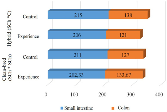
As can be seen from Figure 1, the best indicators of the average linear dimensions of the small and large intestine were in the control group of hybrid breed animals (SCh * C). The most developed colon sections were in the hybrid control group (SCh * C), exceeding the purebred control group (SCh * SCh) by 8% and averaged 138 cm. At the same time, the average linear dimensions of both intestinal sections of purebred rabbits (SCh * SCh) the control group was 211 ± 0.37 and 127 ± 1.67 cm, respectively.
In the groups of animals infested with eimeria, comparative data on the average length of the small intestine were within the statistical error (from 202.33 ± 2.74 to 206 ± 3.65), but the indices in purebred (SCh * SCh) significantly exceeded the SCh * group. C for the thick section, where the average difference was about 13 cm.
The degree of development of the small intestine in the invasive animals of the hybrid group (SCh * C) does not significantly differ in comparison with the control (SCh * C). Still, the large intestine is less developed by 12.3%.
The sensory evaluation of the carcasses did not reveal any visible changes in the quality of the meat.
Pathological studies of internal organs showed that with a low intensity of invasion (50-60 thousand oocysts per head), regardless of the breed of rabbits, pathomorphological changes were found only in the small intestine in the form of small foci of punctate and banded hemorrhages.
During the histological examination of the intestines of both breeds invasive rabbits, despite minor pathomorphological lesions mucous membrane of the small intestine, it has not been finding damage to the enterocytes of the villi and crypts of the unilamellar limb epithelium (Figure 2).
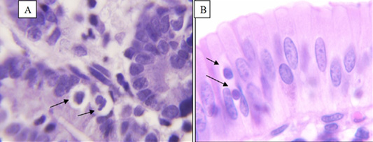
Figure 2: Endogenous stages of eimeria in epithelial cells of the mucous membrane of the small intestine (Coloring: hematoxylin-eosin, magnification: A x400 and B x1000).
In the epithelial plate, endogenous stages of eimeria have been finding at various stages of merogony. Trophozoites oval, round, or drop-shaped with a pronounced basophilic nucleus inside the parasitoform vacuole were find more often in the area of the crypts. Inside some enterocytes, there meronts were consisting of numerous oval-elongated merozoites. Around the parasitic foci were observed blood filling of capillaries, moderate lymphocytosis, and eosinophilia.
The histological examination of the colon, liver, and spleen of the experimental animals did not reveal morphological changes in the structure.
CONCLUSIONS AND RECOMMENDATIONS
Thus, the results of our studies showed that at a low intensity of infection, there is a decrease in weight gain in rabbits of the hybrid breed of the Soviet chinchilla and California (SCh * C) by 25%. However, parasites do not harm the organoleptic characteristics of meat. As a result of measurements of some sections of the length of the digestive tube, it has found that the best indicators of the average linear dimensions of the thin (215 ± 4.38) and thick (138 ± 2.4) intestinal sections were in the animals of the control group of the hybrid breed (SCh * C). The degree of development of the small intestine in the invasive animals of the hybrid group (SCh * C) does not significantly differ in comparison with the control (SCh * C); however, the large intestine is less developed by 12.3%. Histological examination of the organs of the invasive rabbits of both breeds invasive rabbits, despite minor pathomorphological lesions mucous membrane of the small intestine, it has not been finding damage to the enterocytes of the villi and crypts of the unilamellar limb epithelium. The histological examination of the colon, liver, and spleen of the experimental animals did not reveal morphological changes in the structure.
ACKNOWLEDGMENT
The study was carried out with the financial support of the Russian Foundation for Basic Research within the framework of scientific project No 19-316-90059.
AUTHOR’S CONTRIBUTION
Karina Sidorenko was responsible for rabbits feeding and collecting scientific indexes. Manya Mkrtchyan is the scientific leader and developer of this research. Yuri Kuznetsov conducted rabbits infected of experimental groups by Eimeria species E. Perforans and E. irresidua. Ekaterina Klimova was studying the pathogenic effect of these protozoa on animals, its live weight, slaughter weight and slaughter yield of carcasses.
CONFLICT OF INTEREST
The authors have declared no conflict of interest.
REFERENCES


