Advances in Animal and Veterinary Sciences
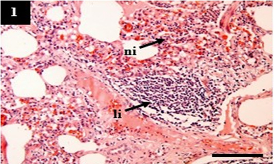
Histopathology of lung from case C1 showed predominantly infiltration of neutrophil (ni) and lymphocytes (li) within the pulmonary interstitial. H&E, 10×.
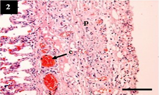
Histopathology of lung from case C4 showed the thickening of the pleural wall (p) and congestion within the venous (c). H&E, 10×.

Histopathology of liver from case C2 showed sinusoidal congestion (c) with a moderate fatty degeneration (fd) in the cytoplasm of hepatocytes. H&E, 40×.
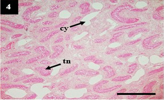
Histopathology of kidney from case C4 showed moderate cystic dilation of tubules (cy) and tubular necrosis (tn). H&E, 4×.
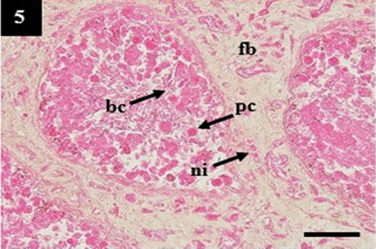
Histopathology of kidney from case C4 showed the deposit of pinkish homogenous (pc) within the lumen that may belong to amyloid, the thickening of fibrous tissue (fb) and infiltration of neutrophil (ni) within the interstitial. H&E, 20×.
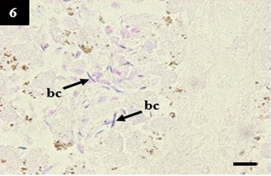
Histopathology of kidney from case C1 showed the slender form bacteria in the lumen that may belong to Leptospira sp. (bc). Gram, 1000×.


