Advances in Animal and Veterinary Sciences
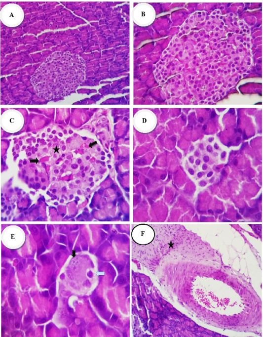
Photomicrographs of the rat’s pancreases of Islet of Langerhans (H and E stain): Group 1 (normal control) showed: (A, B) normal size of islet of Langerhans and Cellularity x200 and x400 respectively. Group 2 (diabetic control) showed: (C) destruction or necrosis of islets (star), congestion of capillaries (thick arrow) with hypocellularity; (D, E) reduction in size, necrosis apoptosis, pyknosis and karyorrhectic of islet cells nuclei (arrows), x400; (F) intense interlobular fibrosis and edema admixed with inflammatory cells and proliferated fibroblasts (star), x200
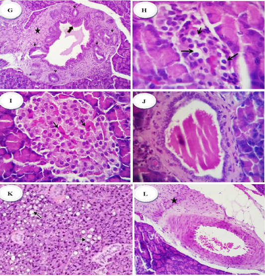
Group 2 (diabetic control) showed hyperplastic epithelium (thick arrow) of commo duct surrounded with chronic inflammatory reaction (mainly lymphocytes and plasma cells) (star) rich in fibroblast (thin arrow), x400 (G) Group 3 (diabetic rats treated with metformin) revealed: (H) reduction in the number of cellular contents especially β-cells with a pyknotic an/or necrotic nuclear changes, x200, Group 4 (diabetic rats treated with ethanolic extract of ginger) showed: (I) normal islets with average number and size of cellular contents, besides mildlu congested capillaries (arrows), (J) mildly dilated pancreatic ducts with retention of the secretory contents, x400. Group 5 (diabetic rats treated with ethanolic extract of N.sativa) showed: (K) islets with pyknotic changes (early necrotic changes), x400 and or vacuolation of the cytoplasm (arrows), (L) perivascular fibrosis (star), x200


