Advances in Animal and Veterinary Sciences
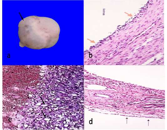
Follicular cyst, 7cm in diameter showing semitransparent membrane (arrow)(a)Histological structure of follicular cyst showing degenerated granulosa cells (arrows)(HEX100)(b)Histological structure of luteal cyst showing vacuolated granulosa cells (arrows)(HEX100)(c)Ovarian hypoplasia, stroma of connective tissue with blood vessels covered with flattened epithelium (arrows)(HEX100)(d).
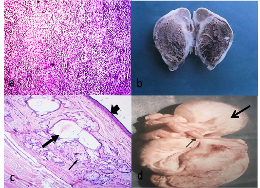
Fibroma, fibroblasts waving in different directions (HEX100)(a)Cystic type of ovarian teratoma about 6.5X4 cm containing hairy material and brownish jelly-like fluid(b) Histological structure of teratoma showing stratified epithelium (arrow head), sweat glands (thick arrow) and sebaceous glands (thin arrow)(HE X40)(c)A complicated case, adhesion (thin arrow), ovarian hydrobursitis (thick arrow) (d).
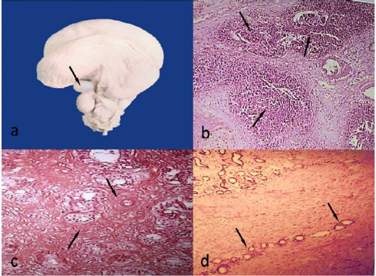
Paraovarian cyst, 1.8cm in diameter (arrow)(a)Acute endometritis, multiple abscesses (arrows) (HEX40)(b)Chronic endometritis, massive fibrosis around glands and blood vessels (arrows)(HEx100)(c)Adenomyosis, uterine glands migrating throughout myometrium (arrows) (HEX40)(d).


