Advances in Animal and Veterinary Sciences
Short Communication
Adv. Anim. Vet. Sci. 8(11): 1180-1183
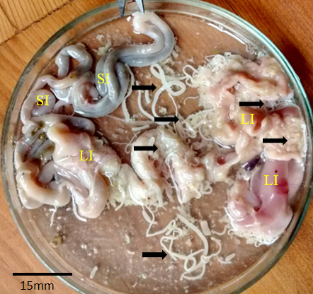
Figure 1
Pigeon intestine with abundance of Raillietina spp. SI: Small intestine; LI: Large intestine; Arrows: Raillietina spp.
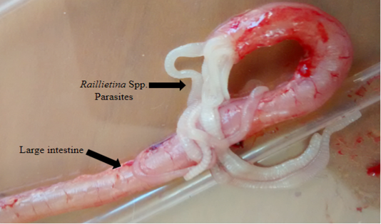
Figure 2
Pigeon large intestine blocked with abundance of Raillietina spp.
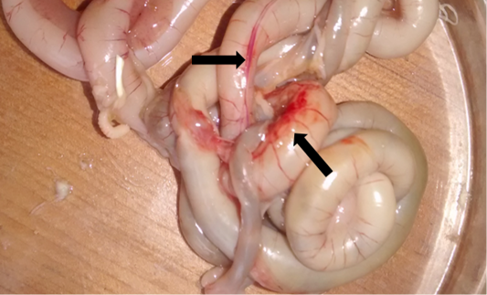
Figure 3
Thickened intestine clearly showing hemorrhage (arrows) at different point.

Figure 4
Raillietina spp. (Fuhrmann, 1920). A: Scolex; B: Immature proglottids; C: Mature proglottids; D: Gravid proglottids occupied by uterine eggs.
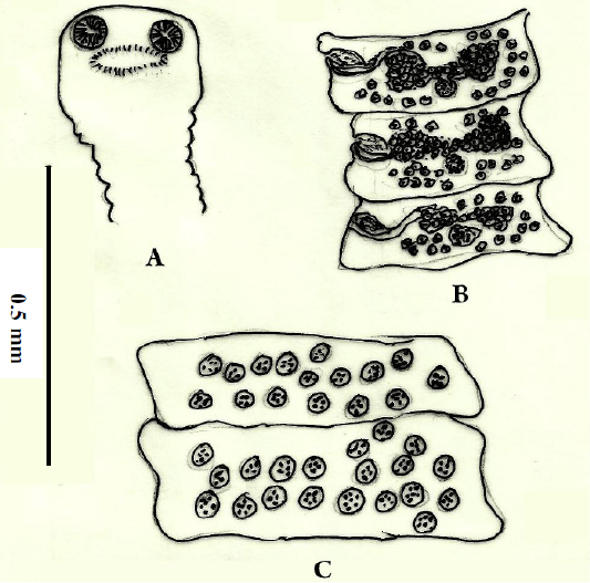
Figure 5
Raillietina spp. (Fuhrmann, 1920). A- Scolex showing suckers and rostellum armed; B- Mature proglottids representing the reproductive organs and the position of cirrus sac; C- Gravid segments occupied by uterine eggs.


