Advances in Animal and Veterinary Sciences
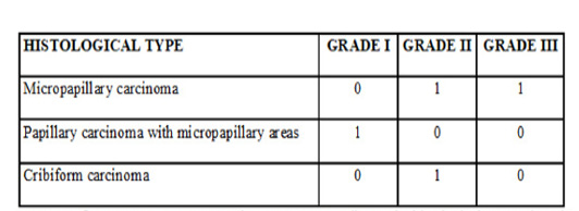
Inflammatory mammary carcinoma cases according to the histological type and grade
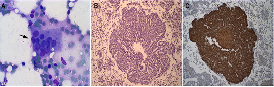
Representative Bone marrow photomicrographs: (A) Epithelial cell clusters in the smear are compatible with DTCs from inflammatory mammary carcinoma (see black arrow). Giemsa staining, obj. 10×. (B) Histopathology of canine bone marrow with the presence of epithelial cells in a cribriform tumor (H-E), obj. 20×. (C) Cytokeratin positive staining in epithelial cells present in canine bone marrow, counterstained in Harri’s hematoxylin, obj. 20×.
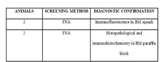
The number of cases, the screening method, and the technique used to confirm the DTCs.
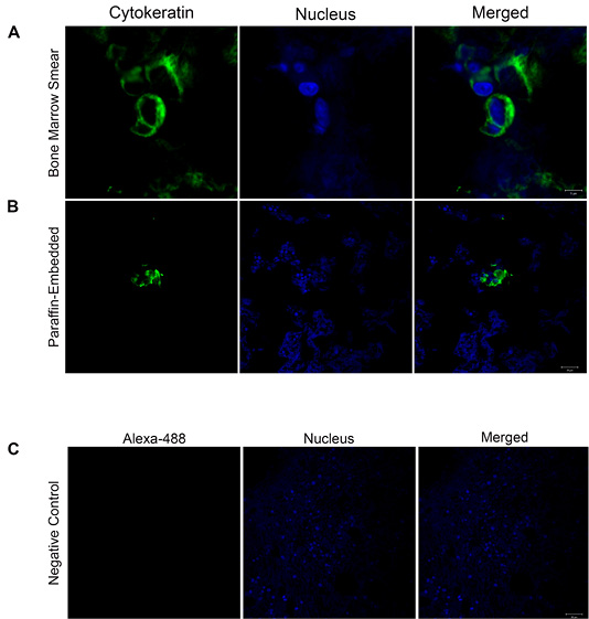
Cytokeratin localization confirmed by confocal microscopy: (A-B) Immunofluorescence images show in green cytokeratin localization in bone marrow smear and paraffin-embedded tissue (Scale Bars= 5 and 20 µm). The nuclei were stained with Hoechst and are shown in blue. The merged images confirm the subcellular localization of cytokeratin. (C) Representative image of the negative control. Images are representative of what was observed in two independent experiments. Scale Bar: 20 µm.


