Advances in Animal and Veterinary Sciences
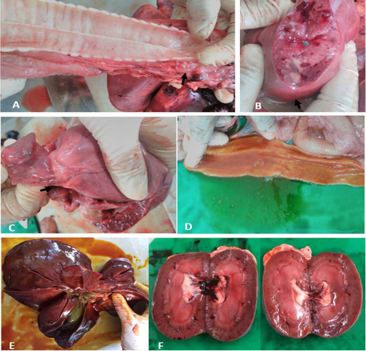
Gross lesions at necropsy. A) Trachea. Frothy exudate in thoracic inlet. B) Lung. Patchy round nodules containing caseous exudate. C) Heart. Whitish spots on the coronary groove of the heart (arrow). D) Intestine. Thicken and redden intestinal mucosa. E) Liver. Enlarged, reddened and firm consistency. F) Kidneys. Generalised reddening of both kidneys.
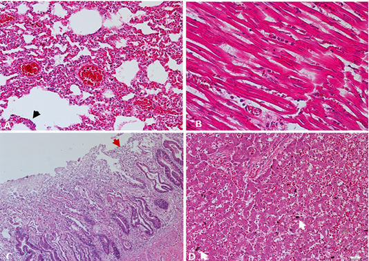
A) Lung. Interstitial pneumonia and pulmonary congestion, including dilatation of capillaries in the alveolar walls filled with red blood cells and thickened alveolar walls with inflammatory cells. Cuboidal hyperplasia of type II pneumocytes in the alveoli was also seen (arrow). H&E. 200X. B) Heart. Lymphocytic myocarditis, degenerated myofibrils with infiltration of macrophages admixed with neutrophils in between cardiac myocytes. H&E. 400X. C) Intestine. Enteritis, degeneration and necrosis of the intestinal villi with mononuclear cells infiltration among enterocytes, with shortened villi (arrow). H&E. 100X. D) Liver. Hepatic congestion, including marked vascular congestion, dilatation of the sinusoids, presence of organizing thrombus and degenerating hepatocytes contains cytoplasmic lipid droplets, with evidence of hemosiderin-laden macrophages (arrow). H&E. 200X.
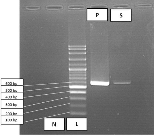
PCR amplification results for Canine Parvovirus in faecal sample. Lane 1, negative control (N); lane 2, 100 bp DNA ladder marker (L); lane 3, positive control (P); lane 4, faecal sample (S) positive for parvovirus at 600 bp.


