Advances in Animal and Veterinary Sciences
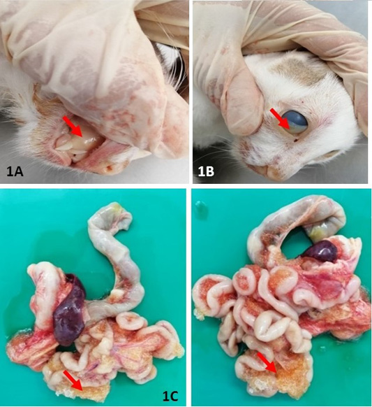
Gross lesion at necropsy: Extensive yellowish discolouration of mucous membrane (A) and conjunctiva (B) were indicative of severe jaundice. (C) and presence of extensive fibrinous plaque on serosal surfaces of intestines. All the lesions described have been indicated in arrow (↘).
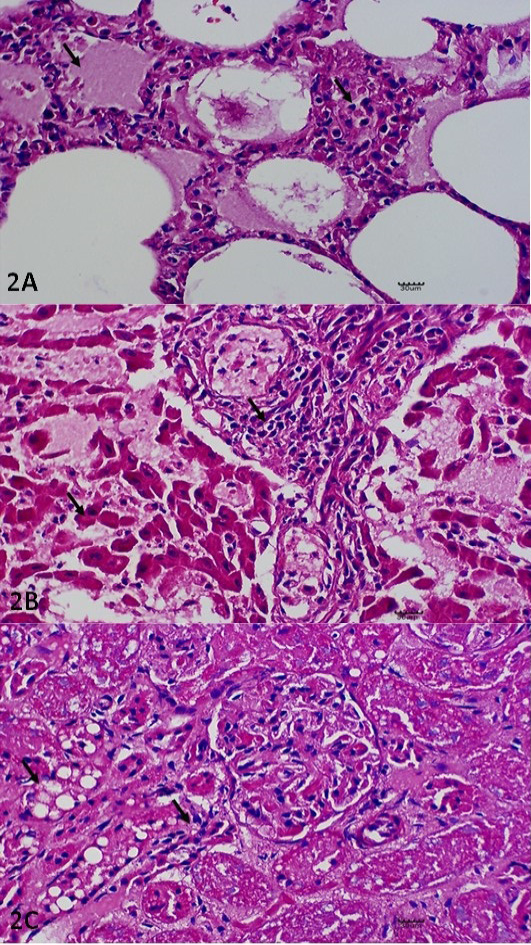
Histopathological Lesions. (A) Pulmonary edema and thickening of the interalveolar septae with leucocytic infiltration. H and E. X400. (B) Necrotising hepatitis, including necrotic changes on the hepatocytes with generalised infiltration of the leucocytes. H and E. X400. (C) Glomerular nephritis, including cytoplasmic vacuolation of the renal tubule (X) and epithelium clusters of scattered necrotic tubules (Y). H and E. X400. All the lesions described have been indicated in arrow (↘).
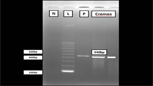
Gel electrophoresis of RT-PCR analysis from Cromox using primers targeting the conserved polymerase gene of FCoV. Lane N: Negative control; Lane L: 100 bp DNA marker (Vivantis, Malaysia); Lane P: Positive control; Lane C: Cromox. Remark: Deionized water was used as negative control.

The unrooted phylogenetic tree between the local isolates FCoV Malaysia Cromox (in text box) with the reference isolates. The phylogenetic tree was constructed using maximum-likelihood method with 1000 bootstrap replicates. Based on this phylogenetic tree, the strain of Cromox is closely related to strain found in China and Netherlands.


