Advances in Animal and Veterinary Sciences
Case Report

Arthrogryposis Multiplex Congentia and Fetal Mummification in a Sheep with Hydrops of Fetal Membranes
Ahmed Saad Ahmed Hassaneen1*, Abdelnaser Abdelmonim Azab2, Islam Farag Fouad1, Mohammed Abdelfattah Noby2, Amany Sayed Mawas3, Nasra Ahmed Yousef1
1Department of Theriogenology, Obstetrics, and Artificial Insemination, Faculty of Veterinary Medicine, South Valley University, 83523 Qena, Egypt, 2Department of Surgery, Anaesthesiology, and Radiology, Faculty of Veterinary Medicine, South Valley University, 83523 Qena, Egypt, 3Department of Pathology, and Clinical Pathology, Faculty of Veterinary Medicine, South Valley University, 83523 Qena, Egypt.
Abstract | Hydrops of the fetal membranes (HFM) and fetal mummification are considered as pathological disorders of pregnant sheep. Arthrogryposis multiplex congentia was reported as a very rare congenital syndrome in newly born/stillbirth lambs or fetuses. A three-years- and sixth-months-old full-term pregnant Rahmani ewe was presented to Veterinary Teaching Hospital, Faculty of Veterinary Medicine, South Valley University, Egypt. The ewe clinically showed HFM and vaginal prolapse, successful delivery revealed 6 dead fetuses; 3 mummified, and 3 full-term malformed fetuses that were characterized by: (1) dysgnathia (brachygnathy inferior), (2) kyphoscoliosis, (3) arthrogryposis, (4) congenital deformities of cervical spine, and (5) abnormalities of the thoracic cage. All these malformations were confirmed by radiographic imaging. The expansion in the use of artificial insemination is strongly required in sheep production in order to minimize the inbreeding-resulted inherited origin diseases such as this case study.
Keywords | Brachygnathy, Ewe, Fetal malformations, Hydrops of fetal membranes, Kyphoscoliosis
Received | April 27, 2020; Accepted | June 21, 2020; Published | July 18, 2020
*Correspondence | Ahmed Saad Ahmed Hassaneen, Department of Theriogenology, Obstetrics, and Artificial Insemination, Faculty of Veterinary Medicine, South Valley University, 83523 Qena, Egypt; Email: ahmed.hassaneen@vet.svu.edu.eg
Citation | Hassaneen ASA, Azab AA, Fouad IF, Noby MA, Mawas AS, Yousef NA (2020). Arthrogryposis multiplex congentia and fetal mummification in a sheep with hydrops of fetal membranes. Adv. Anim. Vet. Sci. 8(8): 848-852.
DOI | http://dx.doi.org/10.17582/journal.aavs/2020/8.8.848.852
ISSN (Online) | 2307-8316; ISSN (Print) | 2309-3331
Copyright © 2020 Hassaneen et al. This is an open access article distributed under the Creative Commons Attribution License, which permits unrestricted use, distribution, and reproduction in any medium, provided the original work is properly cited.
INTRODUCTION
Hydrops of the fetal membranes (HFM) and fetal mummification, as two pathological disorders of pregnant animals, had been reported in different species including sheep, goat and cow. The hydrops of fetal membranes affects the condition of both dam and fetus (Roberts, 2012). Fetal mummification is considered as an uncommon condition in most domestic species including sheep (Lefebvre, 2015). In fetal mummification, fetal death takes place at the middle or last third of gestation without regression of the corpus luteum, and the presence of high progesterone levels lead to retention of the fetus/fetuses with subsequent absorption of fetal fluids resulting in papyraceous mummification with parchment-like fetal membranes (Noakes et al., 2001).
The occurrence of such HFM condition associated with fetal malformations such as arthrogryposis and brachygnathy had been previously reported in sheep, more than forty years ago (Coetzer and Barnard, 1977). Congenital arthrogryposis complicated with musculoskeletal disorders was previously reported in newly born goats (Khodakaram-Tafti et al., 2014). In a recently published report, arthrogryposis was described as a cause of dystocia in sheep (Palanisamy et al., 2018). Moreover, another group described arthrogryposis multiplex congentia (AMC) as a very rare congenital syndrome in lambs (Tejedor et al., 2010). AMC which is characterized by arthrogryposis, kyphoscoliosis was previously found in both lambs and calves (Tejedor et al., 2010; Agerholm et al., 2016).
To our knowledge, there are no available articles that reported the occurrence of the three disorders; HFM, AMC, and fetal mummification altogether in sheep. The aim of the present article is to report and describe such rare novel syndrome of HFM associated with both AMC and fetal mummification in sheep.
Case history and case description
A three-years- and sixth-months-old full-term pregnant Rahmani ewe of (parity 2) was referred to Veterinary Teaching Hospital, Faculty of Veterinary Medicine, South Valley University, Egypt. It showed bilateral symmetrical round-shape abdominal distention with strong straining associated with complete vaginal prolapse appeared as a round red mass (25 cm in diameter) from the rear end of the ewe (Figure 1).
The case had no systemic illness during the whole period of the present pregnancy with a history of giving birth to stillbirth malformed lambs in the previous lambing.
Treatment and radiography
The prolapsed part was thoroughly washed, and reduced as previously described (Hassaneen, 2018). Clinical signs and thoroughly obstetrical examination revealed that the ewe was presented at the time of lambing. After replacement of the prolapsed part, incomplete dilatation of the cervix (ring womb) was detected.
Manual dilatation of the cervix was not successful and a surgical incision was required to widen the incomplete dilated cervical ring. The surgical incision was sutured using simple continuous suture with vicryl suture material after vaginal delivery was completed.
MATERIALS AND METHODS
The clinical and pathological features of the case are fully described below. Radiography was employed to investigate the fetal deformities, the radiographic images of the full term dead fetuses were generated at 10 mAs, 60 kV, and focal distance of 70 cm by the use of a SHIMADZU radiographic device (Model: MD100P, SHIMADZU Corporation, Kyoto, Japan). Medical X-ray films (35 × 43 cm, Arab Co. for Medical Appliances (ACMA), MXB blue, Cairo, Egypt, from MDI, Europa GmbH, Hannover, Germany). While, for radiography images of mummified fetuses; the images were generated at 10 mAs, 50 kV, with the same focal distance of 70 cm using 30 × 40 Medical X-ray films. For more accurate radiographic evaluation of the phenotypic deformities; three orthogonal views were taken for the whole body of the full-term dead fetuses: lateral (L), ventrodorsal (VD), and dorsoventral (DV), however, because of the difficulty of taking the latter two views in the mummified fetuses, only L view was taken. Additional radiographic images were taken for specific parts such as fetal thorax or head in order to make a precise diagnosis. Although, the fetal malformed limbs showed stiffness and limited range of motion, appropriate efforts were made in order to forcibly tract the malformed limbs as possible to avoid superimposition.
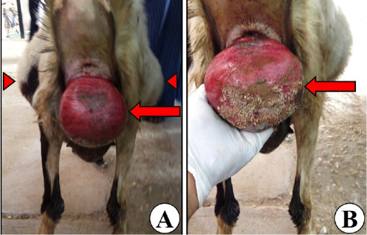
Figure 1: Representative images of the full-term pregnant ewe’s back view showing bilateral symmetrical round-shape abdominal distension as a clinical sign of hydrops of the fetal membranes (arrow heads; A), and vaginal prolapse (arrows; A, B).
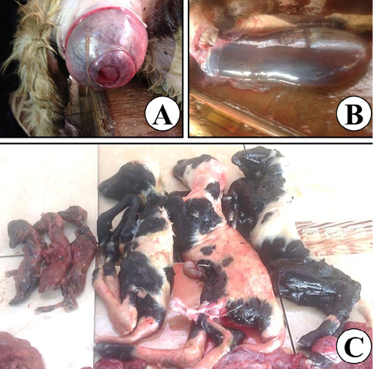
Figure 2: Representative images for the assisted delivery of the affected ewe showing failure of the cervical dilatation (A), distended amniotic sac (B), and 6 dead fetuses; 3 mummified, and 3 full-term malformed fetuses (C).
RESULTS
The assisted delivery revealed 6 dead fetuses; 3 mummified, and 3 full-term malformed fetuses. Rapid protrusions of the distended amniotic sacs surrounding the full-term malformed fetuses were also detected (Figure 2). The average body weight (± SEM) of the mummified fetuses was 144.80 ± 7.10 gm and their average length (± SEM) was 22.00 ± 1.30 cm, while the average birth weight (± SEM) of the full term malformed fetuses was 2.20 ± 0.30 kg, and their average length (± SEM) was 46.20 ± 1.20 cm. Measurement of the exact amounts of both allantoic and amniotic fluids was not possible.
Gross pathology and radiography
The fetal membranes of the full-term dead fetuses showed no gross marked signs of inflammations in the placentomes and intercotyledonary areas, and there were some, grossly observed, congested blackish placentomes, while the fetal membranes of the mummified fetuses showed abnormal brown parchment like changes with sticky and cheesy-like texture (Figure 3).
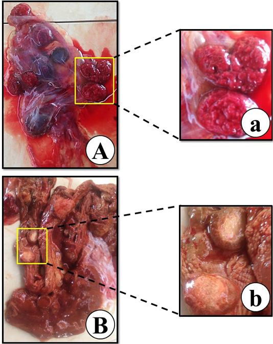
Figure 3: Representative images of the fetal membranes of the full-term dead fetuses (A), abnormal brown parchment like fetal membranes of the mummified fetuses (B), placentomes and intercotyledonary areas of full-term malformed fetuses’ fetal membranes (a), and sticky texture brownish placentomes of mummified fetuses’ fetal membranes (b).
The full term dead fetuses, grossly, showed (1) brachygnathy inferior also named as parrot mouth: as uneven alignment of the upper and lower jaw with abnormal shortness of the mandible, (2) kyphoscoliosis: as a sever curvature of the vertebral column in both longitudinal plane (lateral) (L) view and vertical plane (dorsoventral) (DV) view or ventrodorsal (VD) view, (3) distal arthrogryposis and ankyloses of carpal and tarsal joints, (4) congenital deformities of cervical spine and torticollis which also named as wryneck/twisted neck, and (5) abnormalities of the thoracic cage; ribs and sternum (Figures 4A-C, 5). Even by application of force, correcting the posture of the hyperflexed malformed limbs and twisted neck was not possible due to stiffness of the joints and neck. Ankyloses had been confirmed by the rigidity of malformed joints which had a very limited range of motion. Exact diagnosis of all these deformities had been confirmed by the L, and VD or DV views of radiography (Figure 4).
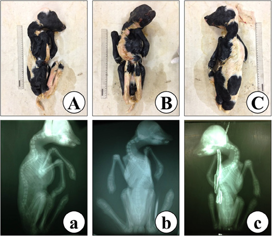
Figure 4: Representative images of a full-term malformed dead fetus malformations, grossly (A-C), and radiography (a-c) showing kyphoscoliosis, ankyloses, brachygnathy inferior, and arthrogryposis in lateral (L) view (A, a), ventrodorsal (VD) view (B, b), and dorsoventral (DV) view (C, c).
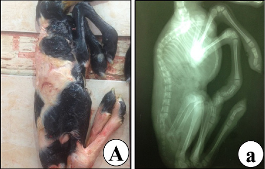
Figure 5: Representative images of a full-term malformed dead fetus showing abnormalities of the thoracic cage thoracic spine, ribs and sternum by lateral (L) view, grossly (A), and radiography (a).
The mummified fetuses appeared grossly as dry mass of brown to black colour with marked kyphosis which had been confirmed by L view radiography (Figure 6).
The heads of mummified fetuses showed brachygnathy superior/undershot as uneven alignment of the upper and lower jaw with abnormal shortness of the maxilla (Figure 7).
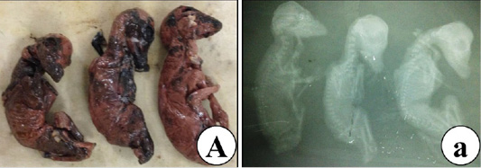
Figure 6: Representative images of lateral (L) view of mummified fetuses showing kyphosis, grossly (A), and radiography (a).
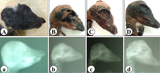
Figure 7: Representative images of lateral view (L) of full-term malformed dead fetus head showing brachygnathy inferior (A, a), and mummified fetuses’ heads showing brachygnathy superior (B-D, b-d), grossly (A-D) and radiography (a-d).
DISCUSSION
This present article fully documented a very rare case of HFM in association with both AMC and fetal mummification in sheep. Moreover, new radiographic findings could be diagnosed not only in the full-term malformed fetuses but also in the mummified ones. Excessive uterine contractions and uterine over-distension were possibly the reasons behind the protrusion of such rounded mass of the vaginal prolapse at the time of lambing.
The dropsical conditions of the fetal sacs have many possible causes including genetic, placental, and fetal hepatic or renal circulation defects (Roberts, 2012). However, the exact etiology of previously reported hydrops-associated congenital malformations was not clear because of the complex mechanisms that may be involved (Coetzer and Barnard, 1977). The same authors had studied the possible association of Rift Valley fever and Wesselsborn disease as etiological agents; this possibility was less likely as all performed serological tests on the affected ewes, lambs or fetuses could not reach a definite conclusion regarding the possible etiological agents.
In our study, the average birth weight of the full-term malformed lambs (BW ± SEM; 2.20 ± 0.30 kg) was lower than that reported in normal Rahmani sheep with multiple-births (BW ± SD; 3.54 ± 1.18 kg) (Gabr et al., 2016). This is because of a possible lower muscle mass (Kalampokas et al., 2012). Although, death of the full-term malformed fetuses before or at the time of lambing is attributed to the malformations of the thoracic cage that involved thoracic spine, ribs and sternum.
The findings reported in this article for the malformed full-term fetuses were largely similar to those defined previously as ovine heritable arthrogryposis multiplex congentia. This previous study found that only one fetus was affected in multiple births (Tejedor et al., 2010). Interestingly, in the present article, brachygnathy inferior was reported in mummified fetuses as a novel deformity in addition to other previously reported deformities. The presence of the deformities in all fetuses would be related with the possible inherited cause.
The presence of the mummified fetuses in association with AMC was helpful to examine such anomalies in the mummified fetuses by the radiographical study. Although dysgnathia was detected in both the full-term fetuses and the mummified fetuses; brachygnathy inferior (overshot/parrot mouth) was detected in the full-term fetuses, while brachygnathy superior (undershot/prognathism) was found in the mummified fetuses. The possible cause of such difference is unknown.
Previous reports support the idea that the cause of the HFM in association with both AMC and fetal mummification reported in this study is more likely due to an autosomal recessive inheritance pattern and associated with inbreeding as previously reported (Nordby et al., 1945; Tejedor et al., 2010; Eriksen et al., 2016). Another report suggested both selenium intoxication and deficiency of manganese as possible causes of kyphoscoliosis and arthrogryposis in cows (Leipold et al., 1974). However, this is less likely to be the cause of the case presented in this article due to absence of any signs of systemic illness either at the time of parturition or even during the whole period of gestation.
The owner was advised to discard this ewe from any further breeding and because natural mating is widely used in sheep reproduction, an eradication strategy should be taken into an account to control this syndrome in sheep.
CONCLUSION
In the present article, HFM that is associated with heritable AMC and fetal mummification was reported in sheep and confirmed by radiographical study. Although, this case is very rare, the inbreeding that is mostly used in sheep farms could increase its incidence due to the possible hereditary cause. Artificial insemination should be widely applied in order to minimize the inbreeding problems especially in the sheep breeds that report this condition.
ACKNOWLEDGMENT
The authors would like to appreciate their deep thanks to Mr. Shaban A. Shahat, Department of Theriogenology, Mr. Sayed A. Mohammed Department of Animal Medicine, and Mr. Mubarak Z. Abdou, Department of Surgery, Faculty of Veterinary Medicine, South Valley University, Egypt, for their assistance, and support.
AUTHORS CONTRIBUTIONS
ASAH, IFF, MAN performed the diagnosis, surgery, radiography and treatment. AAA designed and revised the manuscript. ASM analyzed the pathologic figures and critically revised the manuscript. NAY performed diagnosis, held supportive care and revised the manuscript. ASAH wrote the manuscript. All authors read, revised and approved the final version of the manuscript.
ETHICAL APPROVAL
This article does not contain any studies with human or animal participants. All protocols for diagnosis and treatment followed were in accordance to the Animal Ethics and Use Committee of the South Valley University, Egypt.
Conflict Of Interest
The authors declare that there is no conflict of interest.
REFERENCES


