Advances in Animal and Veterinary Sciences
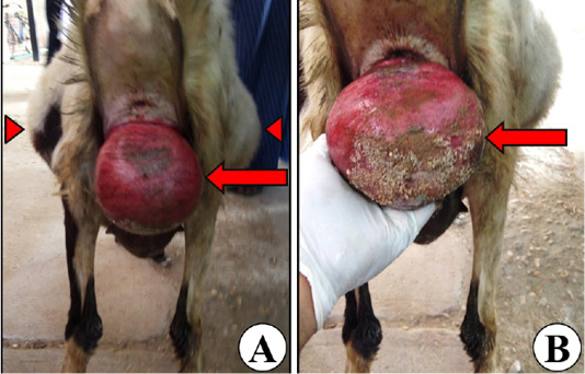
Representative images of the full-term pregnant ewe’s back view showing bilateral symmetrical round-shape abdominal distension as a clinical sign of hydrops of the fetal membranes (arrow heads; A), and vaginal prolapse (arrows; A, B).
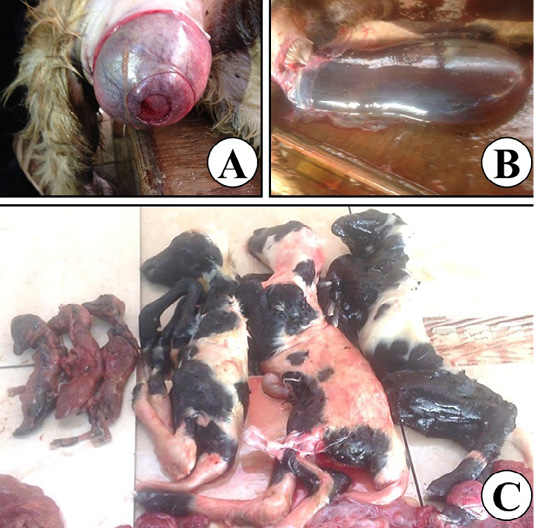
Representative images for the assisted delivery of the affected ewe showing failure of the cervical dilatation (A), distended amniotic sac (B), and 6 dead fetuses; 3 mummified, and 3 full-term malformed fetuses (C).
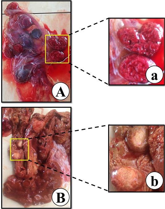
Representative images of the fetal membranes of the full-term dead fetuses (A), abnormal brown parchment like fetal membranes of the mummified fetuses (B), placentomes and intercotyledonary areas of full-term malformed fetuses’ fetal membranes (a), and sticky texture brownish placentomes of mummified fetuses’ fetal membranes (b).
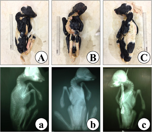
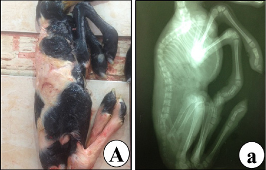
Representative images of a full-term malformed dead fetus malformations, grossly (A-C), and radiography (a-c) showing kyphoscoliosis, ankyloses, brachygnathy inferior, and arthrogryposis in lateral (L) view (A, a), ventrodorsal (VD) view (B, b), and dorsoventral (DV) view (C, c).
Representative images of a full-term malformed dead fetus showing abnormalities of the thoracic cage thoracic spine, ribs and sternum by lateral (L) view, grossly (A), and radiography (a).
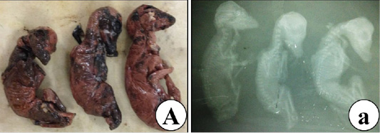
Representative images of lateral (L) view of mummified fetuses showing kyphosis, grossly (A), and radiography (a).
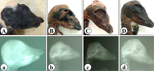
Representative images of lateral view (L) of full-term malformed dead fetus head showing brachygnathy inferior (A, a), and mummified fetuses’ heads showing brachygnathy superior (B-D, b-d), grossly (A-D) and radiography (a-d).


