Advances in Animal and Veterinary Sciences
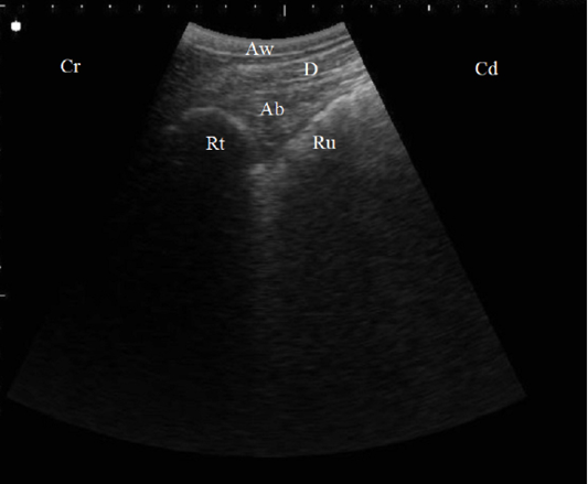
B. Mode ultrasonographic image in a healthy cow. Echogenic half-moon shape reticular wall (Rt) and the cranial dorsal ruminal sac (Ru) imaged from the left 6th ICS, the abomasum (Ab) appeared as a heterogenic structure with echogenic abomasal folds between the reticulum and the diaphragm (D), Aw: Abdominal wall, Cr: Cranial, Cd: Caudal.
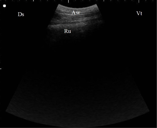
B. Mode ultrasonographic image of the rumen screened from the left flank region in a healthy cow. The ruminal wall (Ru) appeared as an echogenic wall with 0.3 to 0.5 cm in diameter located medial to the abdominal wall (Aw), Ds: Dorsal, Vt: Ventral.
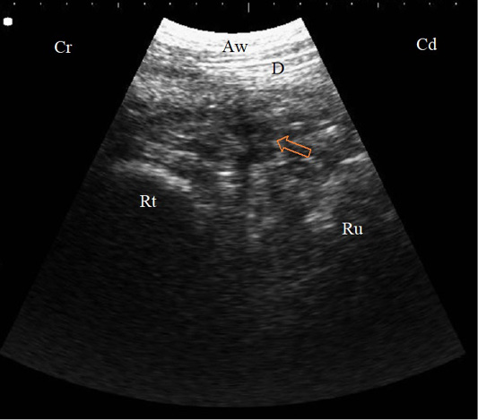
B. Mode sonogram in a case with localized traumatic reticulo-peritonitis. Thickened reticular wall (Rt) separated from the diaphragm (D) with an echogenic fibrinous mass and anechoic exudate (arrow) as imaged from the left 6th ICS, Aw: Abdominal wall, Cr: Cranial, Cd: Caudal.
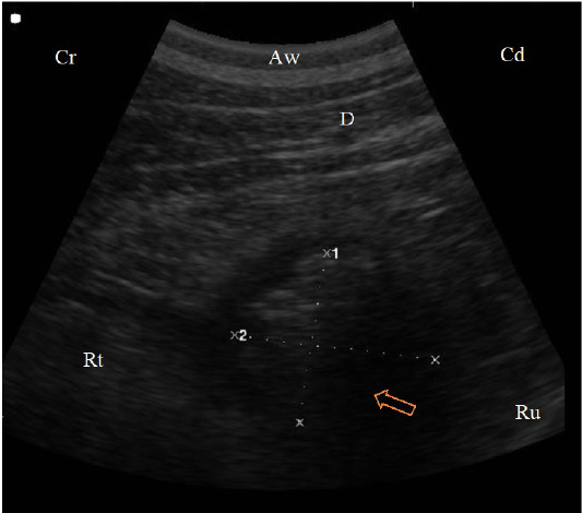
B. Mode sonogram in a case with reticular abscess (arrow) appeared as a circumscribed structure, 4.8 cm diameter, with echogenic wall and anechogenic to hypoechogenic content between the reticulum (Rt) and the diaphragm (D) imaged from the left 7th ICS, Aw: Abdominal wall, Cr: Cranial, Cd: Caudal.
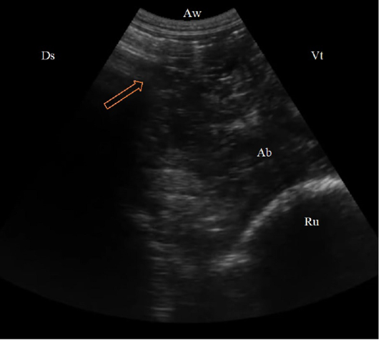
(B. Mode ultrasonographic image in a cow with left abomasal displacement. The abomasum displaced to the left side between the rumen (Ru) and the left abdominal wall as imaged from the left 11th ICS, the reverberation artifacts of the gas cap (arrow) appeared dorsally, followed by the hypoechoic abomasal contents (Ab), Aw: Abdominal wall, Ds: Dorsal, Vt: Ventral.)
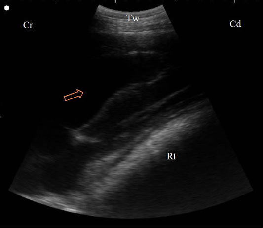
B. Mode sonogram in a case of diaphragmatic hernia imaged from the left 4th ICS. The reticular wall (Rt) compresses the left ventricle of the heart that appeared as echogenic wall with anechoic blood inside (arrow), Tw: Thoracic wall, Cr: Cranial, Cd: Caudal.
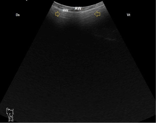
B. Mode ultrasonographic image of ruminal frothy tympany. The small gas gap appeared as reverberation artifact (arrows) medial to the ruminal wall (RW), AW: Abdominal wall, Ds: Dorsal, Vt: Ventral.


