Advances in Animal and Veterinary Sciences
Research Article

Virological Survey of Canine parvovirus Infection in Kars Shepherd Dogs in Kars Province, Turkey
Volkan Yılmaz
Department of Virology, Faculty of Veterinary Medicine, Kafkas University, Kars, Turkey
Abstract | Canine parvovirus type 2 (CPV-2) is responsible for dehydration, severe hemorrhagic enteritis, vomiting and high fever in dogs. This study is a virological investigation of the CPV-2 in Kars Shepherd Dogs owned by small-scale family farming enterprises in the Kars province in Northeast Anatolia, Turkey. Fecal samples were collected from a total of 93 Kars Shepherd Dogs with diarrhea, 48 females and 45 males younger than 1 year of age, unvaccinated for CPV-2. The samples were analyzed for CPV-2 antigens using double antibody sandwich enzyme-linked immunosorbent assay (DAS-ELISA). CPV-2 antigen was detected in 76.3% (71/93), with no significant differences among the genders and age groups (p>0.05). The present study results reveal that CPV-2 infection exists relatively at high levels in Kars Shepherd Dogs. This is the first virological survey for CPV-2 antigens in Kars Shepherd Dogs in the Kars province of Turkey.
Keywords | Canine parvovirus, DAS-ELISA, Dog, Kars, Turkey
Received | April 20, 2020; Accepted | June 04, 2020; Published | June 10, 2020
*Correspondence | Volkan Yılmaz, Department of Virology, Faculty of Veterinary Medicine, Kafkas University, Kars, Turkey; Email: volkankara1980@hotmail.com
Citation | Yılmaz V (2020). Virological survey of Canine parvovirus ınfection in kars shepherd dogs in Kars Province, Turkey. Adv. Anim. Vet. Sci. 8(7): 687-691.
DOI | http://dx.doi.org/10.17582/journal.aavs/2020/8.7.687.691
ISSN (Online) | 2307-8316; ISSN (Print) | 2309-3331
Copyright © 2020 Yılmaz. This is an open access article distributed under the Creative Commons Attribution License, which permits unrestricted use, distribution, and reproduction in any medium, provided the original work is properly cited.
INTRODUCTION
Canine parvovirus type 1 (CPV-1) and Canine Parvovirus type 2 (CPV-2) are types of parvovirus isolated from dogs. CPV-1 was first identified as the cause of mild gastrointestinal and respiratory infections and subacute myocarditis in dogs in 1967, whereas CPV-2 was first identified in the USA in 1978 and, later, was commonly detected worldwide (Berkin et al., 1981; Ozkul et al., 2002; Sakulwira et al., 2003; Yılmaz et al., 2005; Filipov et al., 2011; Gagnon et al., 2016; Miranda et al., 2016). In the 1980s, a new strain of CPV-2 was identified and named CPV-2a, but the virus rapidly mutated and evolved into another strain called CPV-2b in 1984. In addition to the common CPV-2a and CPV-2b that cause death worldwide, CPV-2c, a new and more virulent strain of the virus, was identified in Italy in 2000 (Binn et al., 1970; Parrısh et al., 1991; Buonavoglia et al., 2001). The virus is from the Parvovirus genus of the Parvoviridae family that has non-enveloped, single-stranded DNA and is antigenically related to feline panleukopenia virus. Compared with adult dogs, the infection progresses more severely in puppies and dogs younger than 1 year of age with clinical symptoms of fever, diarrhea, vomiting, severe dehydration, and non-suppurative myocarditis. The disease prognosis is poor due to the dehydration caused by vomiting and enteritis that occur within 24 hours, especially in puppies (Murphy et al., 1999; Decaro and Buonavoglia, 2012; Geetha, 2015). The cardiac syndrome, or myocarditis of CPV-2 can affect pups under 3 months old. This form is rarely seen because infected pups die without clinical symptoms or die shortly after clinical symptoms appear. In addition, 70% pups will die in heart defect by 8 weeks old and the remaining 30% will have pathological changes which may result in death many months or even years later. The most impressive symptom of CPV-2 myocarditis is the sudden death in young pups usually about 4 weeks old (Nandi and Kumar, 2010). Infected dogs shed CPV-2 for 3-12 days in their feces. The primary transmission route of CPV-2 among dogs is fecal-oral. Pathologically, marked duodenal and jejunal villous degeneration with intestinal mucosal and mesenteric lymph node, edema and congestion are noted (Nandi and Kumar, 2010).
CPV-2 infection clinically resembles several other viral infections in dogs, so a definitive diagnosis requires laboratory confirmation and several diagnostic methods, including enzyme linked immunosorbent assay (ELISA), polymerase chain reaction (PCR) analysis, hemagglutination (HA) and electron microscopy (EM) may be used (Pollock and Coyne, 1993; Desario et al., 2005). The double antibody sandwich ELISA test using monoclonal antibodies detects low levels of CPV antigen in feces. It is a rapid, simple, sensitive, and appropriate test for use in the routine diagnosis and epidemiological studies of the CPV-2 antigens in dog feces (Pollock and Coyne, 1993; Prittie, 2004; Desario et al., 2005; Nandi and Kumar, 2010).
Kars Shepherd Dogs are bred in the Northeast part of Turkey, especially in the Kars region. This region of Turkey neighbors other Caucasian countries. Kars Shepherd Dogs are characteristically similar to the Caucasian Ovcharka, present in Georgia, Armenia, Azerbaijan and Iran. Kars Shepherd Dogs are specific Turkish livestock-guarding breed gathered under the general rubric of Turkish shepherd dogs (Yıldırım et al., 2009). Kars Shepherd Dogs were only reported for the first time by Nelson (1996).
Kars Shepherd Dogs are strongly muscled and well boned. The head is large and mastiff and often has a dark fascial mask. Coat color is dark or light agouti gray, and it is lighter (light gray or yellowish) towards the tail and legs. Coat length can be long, medium or short. Ears are triangular and hang tight to the head. Eyes are brown or dark brown, medium-sized and oval (Kırmızıbayrak, 2004).
In the present study, a virological survey for CPV-2 was performed on Kars Shepherd Dogs with diarrhea, owned by small-scale family farming enterprises in the Kars province, using double antibody sandwich ELISA.
MaterIals and Methods
Ethıcs statement
This research was conducted after the approval of Kafkas University Animal Testing Local Ethics Council (Approval Number: KAU- HADYEK-2016-064).
Sample collection
In this study, fecal samples were collected from a total of 93 Kars Shepherd Dogs with diarrhea, owned by small-scale family farming operations in the Kars Province. Of the animals that were sampled in the study, 48 were females and 45 were males (Table 1). Fifty-two of the animals were 1-3 months of age, 16 were aged 3-5 months of age, 15 were 5-7 months of age and 10 were 7-11 months of age (Table 2). None of the dogs were vaccinated against CPV-2, so secondary immunization is not concerned in this study. Collected fecal samples were kept at -80 ºC until analyzed.
Table 1: The distribution of CPV-2 antigen (Ag) positivity according to gender groups.
| Gender | No. of the dogs | CPV-2 | |
| Ag (+) | (%) | ||
| Male | 45 | 38 | 84.4 |
| Female | 48 | 33 | 68.7 |
| Total | 93 | 71 | 76.3 |
Table 2: The distribution of CPV-2 antigen (Ag) positivity according to age groups.
| Age (months) | No. of the dogs | CPV-2 | |
| Ag (+) | (%) | ||
| 1-3 | 52 | 41 | 78.8 |
| 3-5 | 16 | 12 | 75.0 |
| 5-7 | 15 | 11 | 73.3 |
| 7-11 | 10 | 7 | 70.0 |
| Total | 93 | 71 | 76.3 |
The Kars Shepherd Dogs, raised throughout the Kars province, guard herds in the high mountains and valleys of northeast Turkey, and are adapted to local climatic conditions. They have characteristics that are similar to the Caucasian Ovcharkain (Caucasian Mountain Dog) of the Caucasus countries bordering the Kars region. These dogs have a highly developed instinct for protection and protect herds, homes as well as businesses, and therefore are important for the people in this region. In order to ensure the survival of this local breed, which is present in small numbers over a limited geographical area of Turkey, important diseases must be studied and the necessary measures must be taken against these diseases (Yılmaz, 2017). The Kars Shepherd Dogs have mostly been used for shepherd dogs in small-medium scale private goat and sheep breeding enterprises, so number of the dogs per farm was among 1 to 7. Mentioned dogs are native pure breed dogs in Northeast Anatolia but they have no pedigree papers. Because vaccination does not apply to work in these dogs in the region, none of these dogs are vaccinated against any viruses before.
Double antıbody sandwıch elısa (DAS-ELISA)
Commercial DAS-ELISA (Agrolabo, Italy, Cat. No: 27224032), used for the detection of CPV-2 antigens, was carried out according to the manufacturer’s instructions. At the first stage, each of the fecal samples was diluted using Sample and Conjugate-I diluent. Briefly, 100 μL of tested fecal samples were added to wells, and incubated for one hour at 37°C and then washed four times with wash solution (diluted in deionized water). Then, 100 μL of conjugate-I were added to each well and incubated for 60 min at 37°C. Then four times washing, and 100 μL of conjugate-II were added to wells, and incubated at room temperature for 15 min, and then washed four times. Then 100 μL of substrate solution were added to wells, and incubated at room temperature for 10 min, the enzymatic reaction was stopped by adding 100 μL of stop solution. DAS-ELISA results were analyzed with an automated ELISA reader (Epoch, BIO-TEK, USA) at 405 nm. According to the kit procedure: Cut Off value was calculated using controls (Cut Off= OD Positive control × 0.2 ; Cut Off= OD Negative control × 0.2). It was deemed positive if sample with an OD higher than the positive Cut Off and negative if sample with an OD lower than the negative Cut Off.
Statıstıcal analysıs
The chi-square (χ2) test was used to compare the proportion of positive CPV-2 virological results among different genders and age groups (Minitab 14.0 Inc., State College, PA, USA). Significance level was set at P <0.05.
RESULTS and DIscussIon
This study included 93 Kars Shepherd Dogs, aged < 1 year, owned by small-scale family farming enterprises (1 to 7 dogs per operation) in the Kars region, not vaccinated for CPV-2. The CPV-2 antigen was detected in 76.3% (71/93). The gender-based antigen positivity distribution rates were 68.7% (33/48) for females and 84.4% (38/45) for males, with no statistically significant difference between genders (p>0.05) (Table 1). Animals were grouped according to the ages, the highest antigen positive ratio (78.8%) was found in 1-3 months-old dogs. In this study, 41(78.8%) of the 52 animals aged 1-3 months were found to be positive. 12 (75%) of the 16 animals aged 3-5 months were found to be positive. Of the 15 animals aged 5-7 months, 11 (73.3%) and of the 10 animals aged 7-11 months, 7 (70%) were found to be positive. The ratio of the CPV-2 antigen positivity was found to decrease by the age, however there was no statistically significant difference between age groups (p>0.05) (Table 2).
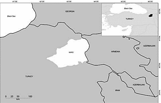
This study was conducted in the Kars province, north-eastern Turkey (43.05° E and 40. 36° N), and its vicinity. It is mountainous and has a cold climate and neighbors other Caucasian countries (Figure 1). Canine Adeno Virus (CAV) was reported for the first time by Yildirim et al. (2009) in Kars Shepherd Dogs in the same region where this study was conducted and detected seropositivity as 65.95%. Canine Corona virus (CCoV) was reported for the first time by Yilmaz (2017) in Kars Shepherd Dogs and detected seropositivity as 75.49%. There was no further extensive investigation in Kars Shepherd Dogs to detect viral diseases in Turkey.
In this study, the antigen positivity rate for CPV-2 in Kars Shepherd Dogs with diarrhea in the Kars region was 76.3% (71/93 dogs). This percentage is higher than the percentages found in studies (Sakulwira et al., 2003; Behera et al., 2015; Timurkan and Oğuzoğlu, 2015; Castro et al., 2017;Yeşilbağ et al., 2017) conducted in past years and is similar to the percentages reported by Miranda et al. (2016). In a study carried out in Thailand (Sakulwira et al., 2003), the presence of CPV-2 in dogs with symptoms of gastroenteritis was 62.8% (44/70) and the infection was more commonly found in 3-6-month-old puppies. A study using PCR by Behera et al. (2015) in India found the CPV rate in dogs was 40.85% (29/71). The distribution of CPV-positive samples by age was 27.59% in 1-3-month-old dogs, 41.37% in 3-6-month-old dogs, 27.59% in 6-12-month-old dogs, and 3.45% in dogs older than 12 months of age. In the same study, the distribution of the CPV-positive samples by gender was 86.21% in male dogs and 13.79% in female dogs. In Brazil, Castro et al. (2017) determined a CPV positive rate of 46% (157/341) in the feces samples collected from 341 dogs with gastroenteritis, younger than 7 months of age, but the researchers did not find a correlation between CPV-positivity and gender or age. Timurkan and Oğuzoğlu (2015) determined the presence of CPV in 25 (38.4%) samples collected from 65 dogs showed the clinical signs of parvovirus infection and sequence analysis revealed that 68% (17/25) contained the CPV2a strain and 32% (8/25) contained the CPV2b strain. In a similar manner, Yeşilbağ et al. (2017) determined a CPV-2 positivity rate of 35.5% in puppies with diarrhea and the characterization study revealed the CPV2a rate was 62.9% and CPV2b rate was 37.1%. In Portugal, Miranda et al. (2016) determined a CPV positivity rate of 76.15% (198/260) in feces samples collected from dogs. In the same study, the prevalence of the CPV variations was 51.5%, 47.5%, and 1% for CPV2c, CPV2b, and CPV2a, respectively.
From the results presented CPV-2 is common in Kars Shepherd Dogs. CPV-2 is highly contagious and spreads rapidly through susceptible dogs. Infected dogs shed virus in their stool in gigantic amounts during the 2 weeks following exposure, fecal contamination of the environment is the important source for virus transmission via ingestion. Because CPV-2 is transmitted feco-orally, it has been determined that the spread of the disease and prevalence rate are particularly high in environments with a large number of dogs in close contact (e.g. dog breeding farms, shelters) (Nandi and Kumar, 2010). In agreement with these, in the present study, the antigen positivity rate was relatively high in family operations, in which several dogs are kept together in close contact, and, with stray dogs roaming free. Furthermore, the relatively higher prevalence of CPV-2 was attributed to bad shelter conditions and irregularity in health controls.
In this study, the samples were collected from 1-11-month-old dogs with diarrhea. Although the results indicated a higher rate of infection in 1-3-month-old dogs, the differences between the age groups were not statistically significant. Despite the results obtained by Aktaş et al. (2011), which showed that age statistically had a significant effect on the disease, the lack of a significant difference in this study was attributed to the non-homogenous age distribution of the dogs from which the samples were collected.
In previous studies (Sakulwira et al., 2003; Behera et al., 2015), the numbers of antigen-positive male dogs were higher than antigen-positive female dogs because of the increased contact between male dogs due to fights for supremacy and competition. Although there was a higher rate of antigen-positivity in male dogs, the difference between the genders was not statistically significant in this study (p>0.05).
In conclusion, the prevalence of CPV-2, in a local dog breed of Turkey called as Kars Shepherd Dogs in the Kars region, Turkey is reported here in for the first time, and is relatively high, indicating that CPV-2 is common among these dogs, and suggesting that preventive measures, such as vaccination, should be performed to decrease CPV-2 associated morbidity and mortality. Furthermore, epidemiological studies of the CPV-2 type are warranted. This study can serve as a reference for molecular characterization studies to determine the antigenic variations of CPV.
Conflict of interest
The authors have declared no conflict of interest.
REFERENCES


