Advances in Animal and Veterinary Sciences
Review Article
Advances in Animal and Veterinary Sciences 1 (4S): 37 – 44Special Issue-4 (Progress in Research on Viruses and Viral Diseases)
Antibody–Based Biosensors for Detection of Veterinary Viral Pathogens
B. Vijayalakshmi Ayyar1, Sushrut Arora2*
- Department of Biochemistry and Molecular Biology, Baylor College of Medicine, One Baylor Plaza, Houston, TX 77030, USA
- Department of Biochemistry and Cell Biology, Rice University, 6100 Main Street, Houston, Texas 77005, USA;
*Corresponding author:drsarora@gmail.com
ARTICLE CITATION:
Ayyar BV and Arora S (2013). Antibody–based biosensors for detection of veterinary viral pathogens. Adv. Anim. Vet. Sci. 1 (4S): 37 – 44.
Received: 2013–11–11, Revised: 2013–12–05, Accepted: 2013–12–07
The electronic version of this article is the complete one and can be found online at
(
http://nexusacademicpublishers.com/table_contents_detail/4/144/html
)
which permits unrestricted use, distribution, and reproduction in any medium, provided the original work is properly cited
ABSTRACT
Improved, cost–effective and rapid diagnostic are highly desirable for detection of veterinary pathogens. They are further desirable for veterinary viral pathogen detection as these pathogens generally cause major ailments in animals. Additionally, there are numerous emerging and re–emerging viral pathogens with many of them being zoonotic, and having public health implications. However, the conventional methodologies for viral detection possess numerous lacunae and, subsequently, they fail to provide the indispensable and timely advantages desired for early diseases intervention. Biosensors offer a lucrative alternative to pathogen detection and their global market is rapidly increasing. Antibody–based biosensors are a class of biosensors with high specificity and have the potential of revolutionizing pathogen detection. They offer numerous advantages over the conventional or molecular methodologies, with the most significant being the option of "on–site” pathogen detection. As of yet, there are limited reports of the application of antibody–based biosensors in veterinary viral detection. However, we feel this technology holds a lot of potential, especially in wake of the recent developments in the areas of antibody–generation, nanotechnology and microfluidics along with the availability of improved antibody immobilization strategies. Consequently, it remains to be seen if biosensors can seize a part of the growing veterinary diagnostics market.
INTRODUCTION
Infectious diseases of animal not only account for economic losses rendered by increased treatment costs, loss of production, morbidity and/or mortality but, additionally, many of these diseases pose public health risk as the pathogens involved harbor the potential of transmission to humans. The risks are further aggravated in case of viral infections which are comparatively difficult to detect and treat. The pathogens involved possess the ability to adapt themselves for survival, by mechanisms such as mutation, recombination, reassortment, infecting new hosts and acclimatizing to new environment.
Viral pathogens are responsible for some of the major infectious diseases of animals (Palmarini, 2007). A number of such diseases, with huge economic repercussions, e.g. classical swine fever (CSF), foot–and–mouth disease (FMD), infectious bursal disease (IBD), bovine virus diarrhea (BVD), canine distemper, swine influenza (SI), chicken infectious anemia (CIA), avian influenza (AI), rinderpest, bluetongue disease, peste–des–petits ruminants (PPR), Newcastle disease (ND), sheep and goat pox, infectious bovine rhinotracheitis (IBR), Marek's disease (MD), pseudorabies, porcine reproductive and respiratory syndrome (PRRS), etc. have been a thorn in the flesh of veterinarians for years now (Balamurugan and Kataria, 2006; Palmarini, 2007; Patel and Heldens, 2009). In addition, recent times have seen emergence of a number of emerging and re–emerging veterinary viral pathogens (Pépin and Tordo, 2010) and a high percentage of them have zoonotic implications (Heeney, 2006).
Rapid Diagnostics – Need of the Hour
Traditional methodologies for viral detection include isolation, in vitro culture, electron microscopy and immunoassays. These methods require specialized technical staff and equipment, and are strenuous, time–consuming, expensive and mostly insensitive (Villarreal, 2010; Dahlhausen, 2010). In addition, conventional assays lack the convenience of "on–site” testing and require a complex work flow starting from sample collection, sample labeling, sample storage and transport to appropriate facility, followed by further sample processing, after which the samples are assayed and the results are interpreted. In the meantime, there is risk of alleviating diseases conditions, spread on infectious disease affecting more population than the initial count and even death due to absence of appropriate disease intervention (Dahlhausen, 2010). Apart from the aforementioned disadvantages, possibilities of variations induced by the personnel, transportation of samples, processing and the testing conditions and lack of uniform analytical platforms further complicates the process making the data unreliable. Additionally, many of the emerging and re–emerging viruses can only be handled at laboratories having recommended biosafety facilities (Poon et al., 2009) which are not very prevalent, even in developed countries. These high risk viruses also present a risk of accidental release while shipping or handling of samples causing further spread and, sometimes, serious public health issues (Poon et al., 2009). Subsequently, there is an impetus for rapid detection of viral pathogens to expedite inferences and, consequently, help in development of prompt measures to arrest the disease progression.
Molecular detection methodologies revolutionized the field of pathogen detection in last few years and have shown better sensitivities than antibody–based assays (Arora et al., 2006). However, these tests though rapid, have their drawbacks. Most of these methods require nucleic acid extraction, skilled personals and equipment, and are costly. The ever growing diagnostic market now emphasizes on generating sensitive and portable assays capable of rapid diagnosis. Modern technologies have made it possible to assemble complex analytical platforms into a single miniature device, biosensors, capable of multitasking from sample processing to detection within seconds to minutes, resulting in rapid turnaround time (TAT). Biosensors are easy to use and do not require trained personnel, laboratory equipment or reagents, thus, capable of testing and yielding results onsite, cutting short the lengthy process.
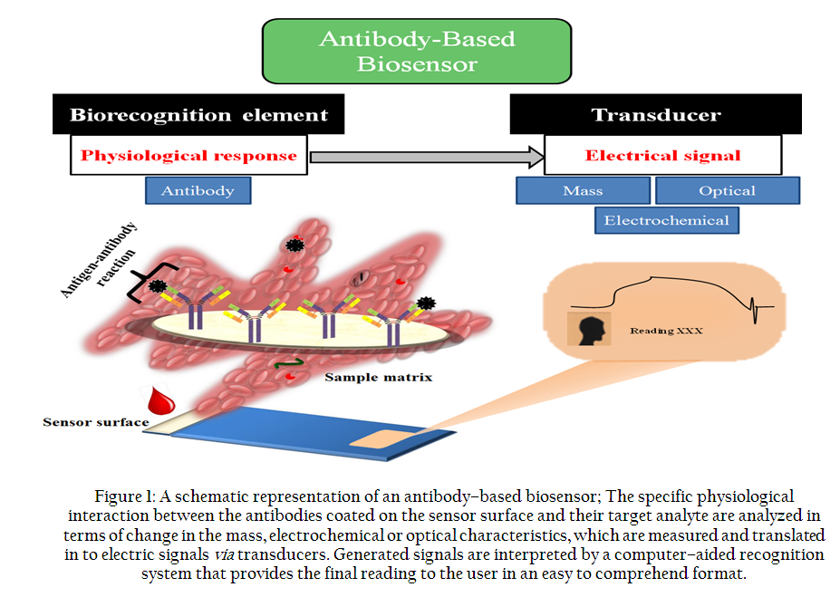
Figure 1: A schematic representation of an antibody–based biosensor; The specific physiological interaction between the antibodies coated on the sensor surface and their target analyte are analyzed in terms of change in the mass
Biosensors
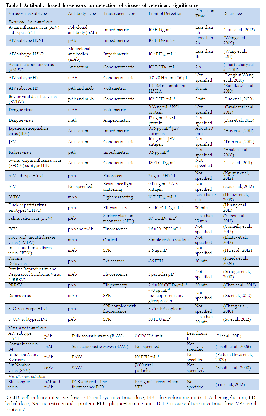
A biosensor is a compact analytical device with a ligand–specific biorecognition element, e.g. antibody, enzyme, receptor, nucleic acid, aptamers, peptide/protein, lectin, cells, tissue or whole organisms, immobilized on a sensor surface integrated directly or indirectly with a signal conversion unit called transducer. The physiological interaction between the ligand and the biorecognition element is translated, by the transducer, into a measurable electric signal, which is further deciphered by a computer–aided readout system for the user (Arora et al., 2010). Biosensors are chiefly classified based on the biorecognition element and the transducers. Antibody–based biosensors employ antibody as biorecognition element (Figure 1) and herein we will be focusing only on them as they are well suited for development of point–of–care–diagnostics and "on–site” diagnostics (Conroy et al., 2009). Consequently, a number of antibody–based biosensors are reported in literature. Table 1 presents some of the recent works on development of antibody–based biosensors for detection of viral pathogens of veterinary significance. Some of these biosensors were developed exclusively to aid in diagnosis of veterinary and zoonotic viruses (Stringer et al., 2008; Luo et al., 2010; Lum et al., 2012), a few were developed for detection of human viral pathogens but can be applied to veterinary diagnostics (Cavalcanti et al., 2012; Tran, 2012), while others were developed as surrogate to similar human viral pathogens (Connelly et al., 2012; Yakes et al., 2013).
Antibodies as Biorecognition Elements
Recent times have witnessed a sudden surge in antibodies applications (discussed by Ayyar et al., 2012), contributed by advances in molecular biological techniques. Due to the simplification of protein expression, peptide synthesis and purification processes, target antigen can be generated in large amounts for antibody production and characterization. Further development of molecular platforms and recombinant DNA technology has aided in selection of high affinity antibodies and tailoring it to desired characteristics. Availability of improved antibody purification methodologies has also contributed to antibody’s increased utilization (Ayyar et al., 2012).
Biorecognition is the key aspect in a biosensor design so it is very essential to choose the biorecognition probe carefully. Antibodies are undoubtedly the most popular class of biorecognition probes owing to their high binding affinity (10–7 to 10–11 M12 Kd) (Connelly and Baeumner, 2012) and explicit target specificity. These characteristics contribute to the sensitivity and specificity of the biosensor to detect the target in complex sample matrices.
Antibodies or immunoglobulins are Y–shaped proteins produced by B–cell as host's immune response to counter antigen. Antibodies bind the target antigen and destroy it in order to protect the host. A typical antibody (immunoglobulin G, IgG) (Figure 2) is composed of a fragment antigen binding (Fab) region and a fragment crystallizable (Fc) region, made up of four polypeptide chains: two heavy chains (50 kDa) and two light chains (25 kDa). These chains are further divided into variable and constant domains based on the sequence variation. A combination of variable heavy (VH) and variable light (VL) chain binds the antigen. The constant domain activates a cascade of events in the immune mechanism to inactivate the antigen. For biosensor application, a suitable host is immunized with the antigen of interest along with the adjuvant to achieve a specific antibody response against the target. Antibodies used as probes can be polyclonal, monoclonal or recombinant (Ayyar et al., 2012).
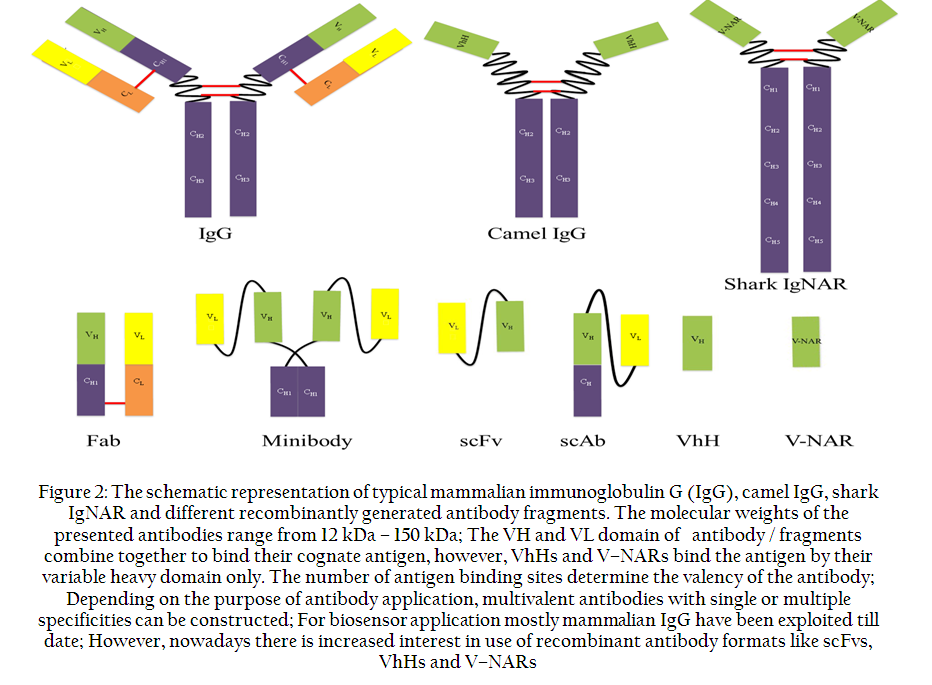
Figure 2: The schematic representation of typical mammalian immunoglobulin G (IgG), camel IgG, shark IgNAR and different recombinantly generated antibody fragments. The molecular weights of the presented antibodies range from 12 kDa – 150 kDa
Polyclonal antibodies (pAbs) are a mixture of antibodies produced against highly immunogenic regions of the antigen with different epitope specificities and clonal affinities, produced by the B cell population of the host in response to an antigen. Large animals such as rabbit, goat and sheep are preferred for pAb generation due to the volume of blood that can be drawn leading to the increased concentration of pAbs harvested from the animal serum. PAb generation is less time consuming and offers the advantages of being stable. PAbs are very useful in detecting similar antigens, contributed by multitude of antibodies obtained against various epitopes of the antigen. However, there are issues of batch variation, lack of high affinity and singular epitope specificity (Ayyar et al., 2012), which can be essential in some of the biosensor applications.
Monoclonal antibodies (mAbs) are obtained by fusion of antibody producing B–cells with the immortal myeloma cells, which leads to the generation of hybrid cells called ‘hybridoma’. Hybridomas retain the antibody–producing feature of B–cells and the immortality processed by the myeloma cells. Each B–cell produces antibody against a single epitope of the antigen, which after rigorous selection and cloning–out procedure are isolated and antibodies are produced from hybridoma cells with single clonal composition. Murine hosts are commonly used for mAb generation, however, there are reports of using rats and rabbits for this purpose. MAbs offers the advantages of being reproducible, large quantity generation, homogeneous composition with high epitope specificity and binding affinity. However, mAb production is expensive and time consuming compared to pAbs and its development requires considerable skills. Recombinant antibodies (rAbs) are antibodies or antibody fragments generated in vitro using molecular techniques. Recombinant antibodies are made by combining the inherent property of immune system along with random recombination of VH and VL, to generate of a vast library of antibodies, which is further screened for specific binders (e.g. Ayyar et al., 2010; Welbeck et al., 2011). RAb generation and isolation is usually time consuming, however, development of large diverse libraries using synthetic and semisynthetic approaches has made it possible to isolate rAbs against various target without going through the tedious immunization and library construction procedures, thus saving time and resources. They are advantageous compared to pAbs and mAbs due to the fact that by employing combinatorial recombinant technologies large library of antibodies can be generated. Antigen specific–selection is done by using molecular display platforms (phage display, ribosome display and yeast display) and further screening based on binding kinetics can be carried out through high–throughput methods (Saerens et al., 2008). Rabs are amenable to engineering for improving its characteristics such as binding, stability, purification and specificity along with the feasibility of format optimization. Discovery of single chain variable fragments (scFv), camelids (VhH) and shark V–NAR, has apprehended much attention in last few years (Saerens et al., 2008; Conroy et al., 2009). These single chain fragments exhibit high stability, expression and purification properties. As biosensor probes they may serve as a better alternative to full length antibodies due to their small size that can help in high density immobilization and reduced non–specific binding due to the absence of Fc fragment (Saerens et al., 2008).
Transducers
A transducer converts the biorecognition event into a measurable signal. Many different types of transducers have been described, however, most of them can be classified as electrochemical, optical, mass–based and calorimetric with the first three being the commonly employed ones for pathogen detection (Luong et al., 2008; Velusamy et al., 2010; Monošík et al., 2012).
Electrochemical biosensors measure the change in electrical properties following biorecognition, as a result of production or consumption of the ions or electrons (Mohanty and Kougianos, 2006). Electrochemical biosensors are further classified into amperometric, potentiometric and impedimetric / conductometric (Velusamy et al., 2010). (i) Amperometric biosensors measure the generated current at a constant potential and are the most commonly used class of electrochemical biosensors (Velusamy et al., 2010). A variant of amperometric biosensors are voltametric biosensors that measure electrical current during controlled variations of the potential. (ii) Potentiometric biosensors measure difference in potential (Velusamy et al., 2010). (iii) Impedimetric / conductometric biosensors function by measuring the change in electrical resistance / conductance of the solution (Mohanty and Kougianos, 2006).
Optical biosensors measure changes in intensity of light. Detection elements in such biosensors are frequently based on luminescence, fluorescence, phosphorescence, colorimetry, reflectance, light polarization and rotation, interference, spectroscopy, ellipsometry and surface plasmon resonance (SPR) (Luong et al., 2008; Velusamy et al., 2010; Huang et al., 2011). However, fluorescence and SPR–based biosensors are most common (Velusamy et al., 2010).
Mass–based biosensors detect a change in mass that occurs following the interaction between the biorecognition element and the target analyte (Mohanty and Kougianos, 2006; Holford et al., 2012). Such sensors generally use piezoelectric materials that change their resonant frequency, following the change in mass, generating acoustic waves. The traveling wave either propagates along the surface of the substrate (surface acoustic wave, SAW) or through the surface of the substrate (bulk acoustic wave, BAW) (Rocha–Gaso et al., 2009). The most commonly used piezoelectric biosensors employ Quartz Crystal Microbalance (QCM) (Holford et al., 2012) and is based on bulk acoustic wave propagation.
Immobilization of Antibodies to the Sensor Surface
Apart from selecting suitable antibodies for probing, a critical factor that influences biosensing mechanism is the immobilization of the antibodies to the sensor surface, which in turn depends on the properties of the sensor interface. Various types of sensor surfaces have been studied (gold, silver, glass, platinum, silica) for this purpose. Ideally sensor surface should be stable, providing large surface area for high density probe immobilization, possessing excellent electrical and thermal conductivity, low diffusion rates, and less signal–to–noise ratio due to matrix effects (Holford et al., 2012). It is hard to get all the desired characteristics on one surface and still keep it small for the device to be portable. Advent of high performance matrices such as carbon nanotubes, fabricated nanoparticles, self–assembled monolayers (SAMs) and quantum dots has led to the development of new generation sensor platforms compatible with the aforementioned sensor needs (Holford et al., 2012). The antibodies can be coupled to the sensor surface by various methods. Passive absorption, covalent coupling, matrix entrapment, encapsulation and affinity tags are the most commonly used methods with their own pros and cons.
Passive Absorption
Physical absorption is a simple process in which the antibodies attach to the sensor surface by Van der Waals, hydrogen or hydrophobic interactions or a combination of all. This process requires minimal antibody manipulation, leaving the antibody unmodified, achieving a high immobilization level. However, as the antibodies are attached by weak interactions, such surfaces have issues of antibody leakage, random antibody orientation, uneven distribution and no tolerance to surface regeneration (Saerens et al., 2008).
Covalent Coupling
Covalent coupling is the most commonly employed method for antibody immobilization to the sensor surfaces. These surfaces are available commercially or can be chemically activated by using glutaraldehyde, periodate, carbodiimide and maleinimides succinimide esters to crosslink the antibodies. Covalent coupling provides a uniform and a stable surface immune to issues like aggregation or antibody leaching over time or regeneration procedures. Conversely, the immobilization requires the manipulation of antibodies, which may render it non–functional or may even degrade it in the process. This can be avoided by protecting the antigen binding region in the antibody before modification (Yoon et al., 2011). General covalent coupling procedures do not ensure proper orientation of the antibodies, which is important for antigen binding and in turn biosensor sensitivity. There are many ways to overcome this issue for e.g. identifying free thiol groups, farther from the active site, or engineering one in the antibodies can help in directional orientation of the antibodies on the gold surface either directly or through a linker or using an intermediate layer such as protein A or G which can be covalently coupled to the surface (Makaraviciute and Ramanaviciene, 2013).
Matrix Entrapment
Matrix entrapment relies on trapping the antibody into a polymeric gel matrix and then immobilizing to the sensor surface. The matrices are thin and porous to allow antigen–antibody interaction. Most commonly used matrices are starch, cellulose, alginate, polyacrylamide, polycarbonate, polyurethane and silica gel. This is a simple and a reliable method of antibody immobilization generating a stable surface, however, it is necessary to ensure that the used matrix is compatible with the sensor surface and will not interfere with antibody interaction (Gupta and Chaudhury, 2007; Monošík et al., 2012).
Affinity Tags
Mostly recombinant antibodies are expressed by conjugating it genetically with peptides / proteins, which acts as an affinity tag, which facilitates its purification and detection. This immobilization technique is based on covalent immobilization of a specific binding partner to the affinity tag on to the sensor surface, creating a stable surface. The chimeric antibody, to be used as biorecognition probe, is passed over the sensor surface causing a directional orientation of the antibody due to affinity tag. Such immobilization procedure requires less/no antibody modification, mild regeneration conditions and the amount of the probe immobilized on the surface can be controlled. This method keeps the binding regions of the probe free and very beneficial in the regard that crude lysate containing antibody can be used, as only specific antibody will be immobilized (Ayyar et al., 2010).
Conclusions and Future Perspectives
It has been more than 50 years since the inception of biosensors (Clark and Lyons, 1962). However, to date only a few biosensors have been commercialized (reviewed by Luong et al., 2008). The main drawbacks that have traditionally prevented the biosensors, in general, from reaching market have been lack of sensitivity, lack of stability and lack of applicability to unprocessed samples (Luong et al., 2008; Monošík et al., 2012). Antibody–based sensors exhibit high specificity owing to the specific interaction, and high affinity and avidity of an antibody with its respective antigen (Saerens et al., 2008; Conroy et al., 2009) and thus, serve as good candidates for use with unprocessed samples. However, in some cases matrix effects may necessitate certain remedial measures (Johnsson et al., 2002; Rodriguez–Mozaz et al., 2006). Most of the reported antibody–based biosensors use polyclonal or monoclonal antibodies (Conroy et al., 2009; Arora et al., 2010) that have stability issues associated to them. In addition, these full antibodies might lose their activity following immobilization on to the sensor surfaces due to disorientation or stearic hindrances (Saerens et al., 2008). However, the availability of chicken antibodies, VhH, V–NAR and different recombinant antibody formats (Figure 2) along with improved immobilization chemistries have helped overcome many of these concerns (Schade et al., 2005; Saerens et al., 2008).
Advances in nanotechnology and microfluidics have allowed miniaturization and subsequently, development of point–of–care and lab–on–a–chip diagnostics (Luong et al., 2008; Wang, 2013). This has revolutionized the field of biosensing and thus, there is increased interest in biosensors. Consequently, there is market expansion in the biosensor market which is expected to be worth by US$16.8 billion by 2018 (http://www.marketresearch.com/Industry–Experts–v3766/Biosensors–Global–Overview–6846583/). However, veterinary pathogen detection is still to harness this technology to its benefit with biosensing currently finding use chiefly in mastitis detection (Viguier et al., 2009).
Viral veterinary diagnostics can greatly benefit from biosensors allowing rapid, robust cheap and simple alternatives to conventional viral detection methodologies. In addition, biosensors allow "on–site” testing, and can be performed and interpreted, within a matter of seconds or minutes, by farmers or veterinarians. This is a lucrative preposition compared to collection and shipment of samples followed by waiting for weeks to get results. Consequently, biosensors can allow veterinarians to provide specific and timely treatment to animals and thus, reducing the resulting morbidity and mortality. Additionally, it allows checking the spread of contagious pathogens to another animals and humans (in case of zoonotic pathogens).
There have been a few studies on development of biosensors for detection of veterinary viral pathogens (Table 1). However, none of the biosensors for veterinary virus detection has made to market yet and it can be rightly concluded that biosensing for veterinary pathogens is still in its infancy. It needs to be seen if the global technology push coupled with the inevitability of cheap, rapid and "on–site” diagnostics in veterinary sector will instigate an upsurge of interest in biosensor development for veterinary pathogen detection.
CONFLICT OF INTEREST
Authors have no conflict of interest to declare.
REFERENCES
Arora S, Agarwal RK and Bist B (2006). Comparison of ELISA and PCR vis–à–vis cultural methods for detecting Aeromonas spp. in foods of animal origin. Int. J. Food Microbiol. 106: 177 – 183.
http://dx.doi.org/10.1016/j.ijfoodmicro.2005.06.019
PMid:16216375
Arora S, Pastorella G, Byrne B, Marsili E and O'Kennedy R (2010). Microbial cells and biosensing: a dual approach–exploiting antibodies and microbial cells as analytical/power systems. In: C.K. Zacharis and P.D. Tzanavaras (eds), Reviews in Pharmaceutical and Biomedical Analysis, Bentham Science Publishers, Sharjah, UAE, 63–75.
Ayyar BV, Arora S, Murphy C and O'Kennedy R (2012). Affinity chromatography as a tool for antibody purification. Methods 56: 116 – 129.
http://dx.doi.org/10.1016/j.ymeth.2011.10.007
PMid:22033471
Ayyar BV, Hearty S and O'Kennedy R (2010). Highly sensitive recombinant antibodies capable of reliably differentiating heart–type fatty acid binding protein from noncardiac isoforms. Anal. Biochem. 407: 165 – 171.
http://dx.doi.org/10.1016/j.ab.2010.07.033
PMid:20696127
Balamurugan V and Kataria JM (2006). Economically important non–oncogenic immunosuppressive viral diseases of chicken–current status. Vet. Res. Commun. 30: 541 – 566.
http://dx.doi.org/10.1007/s11259-006-3278-4
PMid:16883664
Bhatta D, Villalba MM, Johnson CL, Emmerson GD, Ferris NP, King DP and Lowe CR (2012). Rapid detection of foot–and–mouth disease virus with optical microchip sensors. Procedia Chem. 6: 2 – 10.
http://dx.doi.org/10.1016/j.proche.2012.10.124
Bhattacharya M, Hong S, Lee D, Cui T and Goyal SM (2011). Carbon nanotube based sensors for the detection of viruses. Sensors Actuators B: Chem. 155: 67 – 74.
http://dx.doi.org/10.1016/j.snb.2010.11.025
Bisoffi M, Hjelle B, Brown DC, Branch DW, Edwards TL, Brozik SM, Bondu–Hawkins VS and Larson RS (2008). Detection of viral bioagents using a shear horizontal surface acoustic wave biosensor. Biosens. Bioelectron. 23: 1397 – 1403.
http://dx.doi.org/10.1016/j.bios.2007.12.016
PMid:18262781
Cavalcanti IT, Guedes MIF, Sotomayor MDPT, Yamanaka H and Dutra RF (2012). A label–free immunosensor based on recordable compact disk chip for early diagnostic of the dengue virus infection. Biochem. Eng. J. 67: 225 – 230.
http://dx.doi.org/10.1016/j.bej.2012.06.016
Chang Y, Wang S, Huang JC, Su, L, Yao L, Li Y, Wu S, Chen YA, Hsieh J, and Chou C (2010). Detection of swine–origin influenza A (H1N1) viruses using a localized surface plasmon coupled fluorescence fiber–optic biosensor. Biosens. Bioelectron. 26: 1068 – 1073.
http://dx.doi.org/10.1016/j.bios.2010.08.060
PMid:20855191
Chen Y, Huang C, Hou C, Huo D and Jin G (2013). Rapid and label–free detection of porcine reproductive and respiratory syndrome virus on nanoscale by biosensor based on imaging ellipsometry. Integrated Ferroelectr. 145: 122 – 129.
http://dx.doi.org/10.1080/10584587.2013.788963
Clark LC and Lyons C (1962). Electrode systems for continuous monitoring in cardiovascular surgery. Ann. N. Y. Acad. Sci. 102: 29 – 45.
http://dx.doi.org/10.1111/j.1749-6632.1962.tb13623.x
PMid:14021529
Connelly J and Baeumner A (2012). Biosensors for the detection of waterborne pathogens. Anal. Bioanal. Chem. 402: 117 – 127.
http://dx.doi.org/10.1007/s00216-011-5407-3
PMid:21956262
Connelly J, Kondapalli S, Skoupi M, Parker JL, Kirby B and Baeumner A (2012). Micro–total analysis system for virus detection: microfluidic pre–concentration coupled to liposome–based detection. Anal. Bioanal. Chem. 402: 315 – 323.
http://dx.doi.org/10.1007/s00216-011-5381-9
PMid:21909662
Conroy PJ, Hearty S, Leonard P and O'Kennedy RJ (2009). Antibody production, design and use for biosensor–based applications. Semin. Cell Dev. Biol. 20: 10 – 26.
http://dx.doi.org/10.1016/j.semcdb.2009.01.010
PMid:19429487
Dahlhausen B (2010). Future Veterinary Diagnostics. J. Exot. Pet. Med. 19: 117 – 132.
http://dx.doi.org/10.1053/j.jepm.2010.05.006
Dias AC, Gomes–Filho SL, Silva MM and Dutra RF (2013). A sensor tip based on carbon nanotube–ink printed electrode for the dengue virus NS1 protein. Biosens. Bioelectron. 44: 216 – 221.
http://dx.doi.org/10.1016/j.bios.2012.12.033
PMid:23428736
Gupta R and Chaudhury NK (2007). Entrapment of biomolecules in sol–gel matrix for applications in biosensors: problems and future prospects. Biosens. Bioelectron.22: 2387 – 2399.
http://dx.doi.org/10.1016/j.bios.2006.12.025
PMid:17291744
Heeney JL (2006). Zoonotic viral diseases and the frontier of early diagnosis, control and prevention. J. Intern. Med. 260: 399 – 408.
http://dx.doi.org/10.1111/j.1365-2796.2006.01711.x
PMid:17040245
Heinze BC, Song J, Lee C, Najam A and Yoon J (2009). Microfluidic immunosensor for rapid and sensitive detection of bovine viral diarrhea virus. Sensors Actuators B: Chem. 138: 491 – 496.
http://dx.doi.org/10.1016/j.snb.2009.02.058
Hnaien M, Diouani MF, Helali S, Hafaid I, Hassen WM, Renault NJ, Ghram A and Abdelghani A (2008). Immobilization of specific antibody on SAM functionalized gold electrode for rabies virus detection by electrochemical impedance spectroscopy. Biochem. Eng. J. 39: 443 – 449.
http://dx.doi.org/10.1016/j.bej.2007.09.018
Holford TR, Davis F, Higson SP (2012). Recent trends in antibody based sensors. Biosens. Bioelectron. 34: 12 – 24.
http://dx.doi.org/10.1016/j.bios.2011.10.023
PMid:22387037
Hu J, Li W, Wang T, Lin Z, Jiang M, Hu F (2012). Development of a label–free and innovative approach based on surface plasmon resonance biosensor for on–site detection of infectious bursal disease virus (IBDV). Biosens. Bioelectron. 31: 475 – 479.
http://dx.doi.org/10.1016/j.bios.2011.11.019
PMid:22138467
Huang C, Li J, Tang Y, Chen Y and Jin G (2011). Detection of duck hepatitis virus serotype1 by biosensor based on imaging ellipsometry. Curr. Appl. Phys 11: 353 – 357.
http://dx.doi.org/10.1016/j.cap.2010.08.004
http://dx.doi.org/10.1007/s00340-011-4672-3
Huy TQ, Hanh NT, Thuy NT, Chung PV, Nga PT, Tuan MA (2011). A novel biosensor based on serum antibody immobilization for rapid detection of viral antigens. Talanta 86: 271 – 277.
http://dx.doi.org/10.1016/j.talanta.2011.09.012
PMid:22063541
Johnsson L, Baxter GA, Crooks SRH, Brandon DL and Elliott CT (2002). Reduction of sample matrix effects – the analysis of benzimidazole residues in serum by immunobiosensor. Food Agric. Immunol. 14: 209 – 216.
http://dx.doi.org/10.1080/09540100220145000a
Kamikawa TL, Mikolajczyk MG, Kennedy M, Zhang P, Wang W, Scott DE and Alocilja EC (2010). Nanoparticle–based biosensor for the detection of emerging pandemic influenza strains. Biosens. Bioelectron. 26: 1346 – 1352.
http://dx.doi.org/10.1016/j.bios.2010.07.047
PMid:20729069
Lee D, Chander Y, Goyal SM and Cui T (2011). Carbon nanotube electric immunoassay for the detection of swine influenza virus H1N1. Biosens. Bioelectron. 26: 3482 – 3487.
http://dx.doi.org/10.1016/j.bios.2011.01.029
PMid:21354779
Li D, Wang J, Wang R, Li Y, Abi–Ghanem D, Berghman L, Hargis B and Lu H (2011). A nanobeads amplified QCM immunosensor for the detection of avian influenza virus H5N1. Biosens. Bioelectron. 26: 4146 – 4154.
http://dx.doi.org/10.1016/j.bios.2011.04.010
PMid:21536419
Lum J, Wang R, Lassiter K, Srinivasan B, Abi–Ghanem D, Berghman L, Hargis B, Tung S, Lu H and Li Y (2012). Rapid detection of avian influenza H5N1 virus using impedance measurement of immuno–reaction coupled with RBC amplification. Biosens. Bioelectron. 38: 67 – 73.
http://dx.doi.org/10.1016/j.bios.2012.04.047
PMid:22647532
Luo Y, Nartker S, Miller H, Hochhalter D, Wiederoder M, Wiederoder S, Setterington E, Drzal LT and Alocilja EC (2010). Surface functionalization of electrospun nanofibers for detecting E. coli O157:H7 and BVDV cells in a direct–charge transfer biosensor. Biosens. Bioelectron. 26: 1612 – 1617.
http://dx.doi.org/10.1016/j.bios.2010.08.028
PMid:20833013
Luong JH, Male KB and Glennon JD (2008). Biosensor technology: Technology push versus market pull. Biotechnol. Adv. 26: 492 – 500.
http://dx.doi.org/10.1016/j.biortech.2007.07.056
PMid:17826987
Makaraviciute A and Ramanaviciene A (2013). Site–directed antibody immobilization techniques for immunosensors. Biosens. Bioelectron. 50: 460 – 471.
http://dx.doi.org/10.1016/j.bios.2013.06.060
PMid:23911661
Mohanty SP and Kougianos E (2006). Biosensors: a tutorial review. IEEE Potentials 25: 35 – 40.
http://dx.doi.org/10.1109/MP.2006.1649009
Monošík R, Streďanský M and Šturdík E (2012). Biosensors – classification, characterization and new trends. Acta Chim. Slov. 5: 109 – 120.
Nguyen BT, Koh G, Lim HS, Chua AJ, Ng MM and Toh CS (2009). Membrane–based electrochemical nanobiosensor for the detection of virus. Anal. Chem. 81: 7226 – 7234.
http://dx.doi.org/10.1021/ac900761a
PMid:19663392
Palmarini M (2007). A Veterinary Twist on Pathogen Biology. PLoS Pathog. 3: e12.
http://dx.doi.org/10.1371/journal.ppat.0030012
PMid:17319740 PMCid:PMC1803002
Patel JR and Heldens JG (2009). Immunoprophylaxis against important virus diseases of horses, farm animals and birds. Vaccine 27: 1797 – 1810.
http://dx.doi.org/10.1016/j.vaccine.2008.12.063
PMid:19402200
Peduru Hewa TM, Tannock GA, Mainwaring DE, Harrison S and Fecondo JV (2009). The detection of influenza A and B viruses in clinical specimens using a quartz crystal microbalance. J. Virol. Methods 162: 14 – 21.
http://dx.doi.org/10.1016/j.jviromet.2009.07.001
PMid:19628008
Pépin M and Tordo N (2010). Foreword. Vet. Res. 41: 69.
http://dx.doi.org/10.1051/vetres/2010044
PMid:21188838 PMCid:PMC2939695
Pineda MF, Chan LLY, Kuhlenschmidt T, Choi CJ, Kuhlenschmidt M and Cunningham BT (2009). Rapid specific and label–free detection of porcine rotavirus using photonic crystal biosensors. IEEE Sens. J. 9: 470 – 477.
http://dx.doi.org/10.1109/JSEN.2009.2014427
Poon LL, Chan KH, Smith GJ, Leung CS, Guan Y, Yuen KY and Peiris JS (2009). Molecular detection of a novel human influenza (H1N1) of pandemic potential by conventional and real–time quantitative RT–PCR assays. Clin Chem. 55: 1555 – 1558.
http://dx.doi.org/10.1373/clinchem.2009.130229
PMid:19439731
Tran QH, Nguyen THH, Mai AT, Nguyen TT, Vu QK and Phan TN (2012). Development of electrochemical immunosensors based on different serum antibody immobilization methods for detection of Japanese encephalitis virus. Adv. Nat. Sci.: Nanosci. Nanotechnol. 3: 015012.
Rocha–Gaso MI, March–Iborra C, Montoya–Baides A and Arnau–Vives A (2009). Surface generated acoustic wave biosensors for the detection of pathogens: a review. Sensors 9: 5740 – 5769.
http://dx.doi.org/10.3390/s90705740
http://dx.doi.org/10.3390/s9095740
Rodriguez–Mozaz S, Lopez de Alda MJ and Barceló D (2006). Biosensors as useful tools for environmental analysis and monitoring. Anal Bioanal. Chem. 386: 1025 – 1041.
http://dx.doi.org/10.1007/s00216-006-0574-3
PMid:16807703
Wang R, Li Y, Mao X, Huang T and Lu H (2010). Magnetic bio–nanobeads and nanoelectrode based impedance biosensor for detection of avian influenza virus. . In: Nano/Molecular Medicine and Engineering (NANOMED), 2010, IEEE 4th International Conference, 214 – 217.
Saerens D, Huang L, Bonroy K and Muyldermans S (2008). Antibody fragments as probe in biosensor development. Sensors 8: 4669 – 4686.
http://dx.doi.org/10.3390/s8084669
PMCid:PMC3705465
Schade R, Calzado EG, Sarmiento R, Chacana PA, Porankiewicz–Asplund J and Terzolo HR (2005). Chicken egg yolk antibodies (IgY–technology): a review of progress in production and use in research and human and veterinary medicine. Altern. Lab. Anim. 33: 129 – 154.
PMid:16180988
Stringer RC, Schommer S, Hoehn D and Grant SA (2008). Development of an optical biosensor using gold nanoparticles and quantum dots for the detection of porcine reproductive and respiratory syndrome virus. Sensors Actuators B: Chem. 134: 427 – 431.
http://dx.doi.org/10.1016/j.snb.2008.05.018
Su LC, Chang CM, Tseng YL, Chang YF, Li YC, Chang YS and Chou C (2012). Rapid and highly sensitive method for influenza A (H1N1) virus detection. Anal. Chem. 84: 3914 – 3920.
http://dx.doi.org/10.1021/ac3002947
PMid:22401570
Nguyen TH, Ung TDT, Vu TH, Tran TKC, Dong VQ, Dinh DK and Nguyen QL (2012). Fluorescence biosensor based on CdTe quantum dots for specific detection of H5N1 avian influenza virus. Adv. Nat. Sci.: Nanosci. Nanotechnol. 3: 035014.
Velusamy V, Arshak K, Korostynska O, Oliwa K and Adley C (2010). An overview of foodborne pathogen detection: in the perspective of biosensors. Biotechnol. Adv. 28: 232 – 254.
http://dx.doi.org/10.1016/j.biotechadv.2009.12.004
PMid:20006978
Viguier C, Arora S, Gilmartin N, Welbeck K and O'Kennedy R (2009). Mastitis detection: current trends and future perspectives. Trends Biotechnol. 27: 486 – 493.
http://dx.doi.org/10.1016/j.tibtech.2009.05.004
PMid:19616330
Villarreal L (2010). Diagnosis of infectious bronchitis: an overview of concepts and tools. Rev. Bras. Cienc. Avic. 12: 111 – 114.
http://dx.doi.org/10.1590/S1516-635X2010000200006
Wang R, Lin J, Lassiter K, Srinivasan B, Lin L, Lu H, Tung S, Hargis B, Bottje W, Berghman L and Li Y (2011). Evaluation study of a portable impedance biosensor for detection of avian influenza virus. J. Virol. Methods 178: 52 – 58.
http://dx.doi.org/10.1016/j.jviromet.2011.08.011
PMid:21872621
Wang R, Wang Y, Lassiter K, Li Y, Hargis B, Tung S, Berghman L and Bottje W (2009). Interdigitated array microelectrode based impedance immunosensor for detection of avian influenza virus H5N1. Talanta 79: 159 – 164.
http://dx.doi.org/10.1016/j.talanta.2009.03.017
PMid:19559858
Wang T (2013). Micro and nanotechnologies enhanced biomolecular sensing. Biosensors 3: 283 – 285.
http://dx.doi.org/10.3390/bios3030283
Welbeck K, Leonard P, Gilmartin N, Byrne B, Viguier C, Arora S and O'Kennedy R (2011). Generation of an anti–NAGase single chain antibody and its application in a biosensor–based assay for the detection of NAGase in milk. J. Immunol. Methods 364: 14 – 20.
http://dx.doi.org/10.1016/j.jim.2010.09.019
PMid:20883698
Xu J, Wan JW, Yang ST, Zhang SF, Xu N, Li N, Li JP, Wang HY, Bai X and Liu WS. 2012. A surface plasmon resonance biosensor for direct detection of the rabies virus. Acta Vet. Brno 81: 107 – 111.
http://dx.doi.org/10.2754/avb201281020107
Yakes BJ, Papafragkou E, Conrad SM, Neill JD, Ridpath JF, Burkhardt W 3rd, Kulka M and Degrasse SL (2013). Surface plasmon resonance biosensor for detection of feline calicivirus, a surrogate for norovirus. Int. J. Food Microbiol. 162: 152 – 158.
http://dx.doi.org/10.1016/j.ijfoodmicro.2013.01.011
PMid:23416550
Yin HQ, Jia MX, Yang S, Jing PP, Wang R and Zhang JG (2012). Development of a highly sensitive gold nanoparticle probe–based assay for bluetongue virus detection. J. Virol. Methods 183: 45 – 48.
http://dx.doi.org/10.1016/j.jviromet.2012.03.027
PMid:22483982
Yoon M, Hwang HJ and Kim JH (2011). Immobilization of antibodies on the self–assembled monolayer by antigen–binding site protection and immobilization kinetic control. J. Biomed. Sci. Eng. 4: 242 – 247.
http://dx.doi.org/10.4236/jbise.2011.44033
Zou X, Huang H, Gao Y and Su X (2012). Detection of avian influenza virus based on magnetic silica nanoparticles resonance light scattering system. Analyst 137: 648 – 653.
http://dx.doi.org/10.1039/c1an16041a
PMid:22140679


