Advances in Animal and Veterinary Sciences
Case Report
Adv. Anim. Vet. Sci. 8(4): 408-411
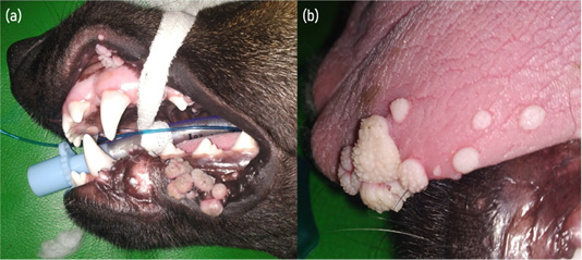
Figure 1
(a) Gross appearance of nodular masses having different sizes present in the lips and gingiva. (b) Multiple nodular mass present at the tongue margin.
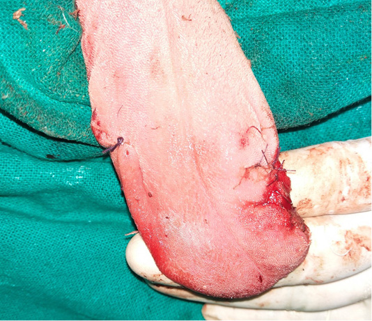
Figure 2
Reconstructed tongue margins following the surgical excision of nodular cauliflower shaped nodules.
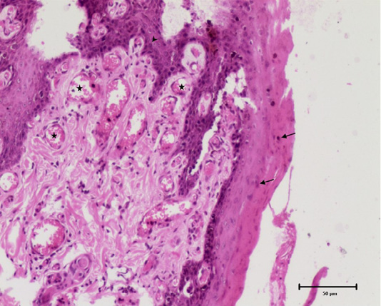
Figure 3
Histomicrograph showing diffuse epidermal hyperplasia with parakeratosis (arrows), broad and elongated rete pegs (arrowhead) and neovascularization of superficial dermis (star). H and E, 10x.
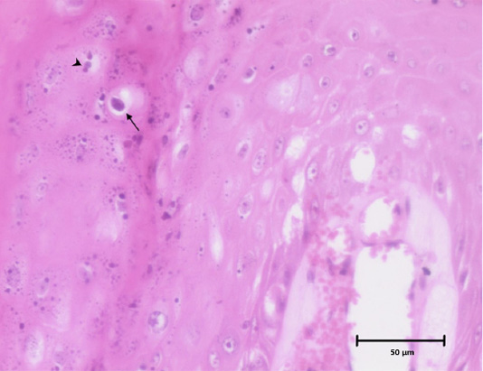
Figure 4
Histomicrograph showing koilocyte with a clear perinuclear cytoplasmic vacuolization (arrow) and clumps of keratohyalin granules (arrow head) in stratum granulosum of epidermis. H and E, 10x.


