Advances in Animal and Veterinary Sciences

Photomicrographs of normal kidney sections showing glomeruli (GL), proximal convoluted tubule (PX), and distal convoluted tubules (DT) (Photomicrographs 1a and 1b). (H and E X 400).
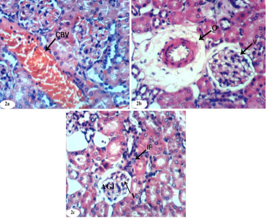
Photomicrographs of kidney sections of doxorubicin-injected group showing dilation and congestion (CBV) of renal blood vessel (Photomicrographs 2a), vacuolation (V) of endothelial lining glomerular tuft, perivascular oedema (O) (Photomicrograph 2b), peritubular mononuclear inflammatory cells infiltration (IF) and atrophied glomeruli (Photomicrograph 2c). (H and E X 400).
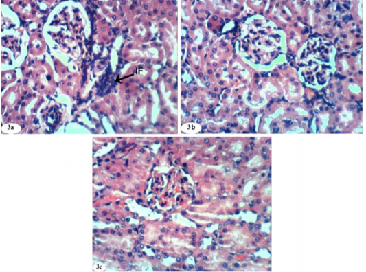
Photomicrographs of kidney sections of doxorubicin-injected group treated with rutin showing mild focal mononuclear inflammatory cells infiltration (IF) (Photomicrograph 3a), improvement and nearly normal structure of the renal tissue (Photomicrographs 3b and 3c). (H and E X 400).

Photomicrographs of kidney sections of doxorubicin-injected group treated with quercetin showing mild congestion (CBV) of intertubular blood vessels (Photomicrograph 4a), vacuolation (V) of endothelial lining glomerular tuft (Photomicrograph 4b) and nearly normal structure of the renal tissue (Photomicrograph 4c). (H and E X 400).
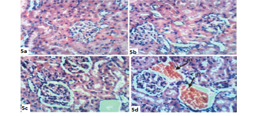
Photomicrographs of kidney sections of doxorubicin-injected group treated with combination of rutin and quercetin showing improvement and nearly normal structure of the renal tissue (Photomicrographs 5a,5b and 5c) and congestion (CBV) of renal blood vessels (Photomicrograph 5d). (H and E X 400).
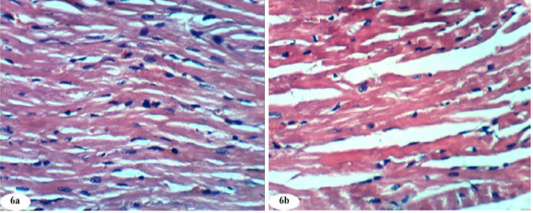
Photomicrographs of normal heart section showed normal histological architecture (photomicrographs 6a and 6b). (H and E X 400).
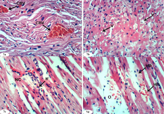
Photomicrographs of heart sections of doxorubicin-injected group showing congestion (CBV) of myocardial blood vessels (Photomicrograph 7a), vacuolization (V) of sarcoplasm of cardiac myocytes (Photomicrograph 7b), focal necrosis (NC) of cardiac myocytes, inflammatory cells’ infiltration (Photomicrograph 7c), intermuscular oedema (O) and haemorrhage (HR) (Photomicrograph 7d). (H and E X 400).
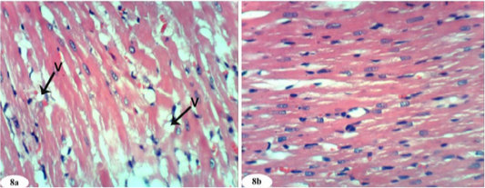
Photomicrographs of heart sections of doxorubicin-injected group treated with rutin showing vacuolization (V) of sarcoplasm of cardiac myocytes (Photomicrograph 8a) and improvement of the tissue in some animals (photomicrograph 8b). (H and E X 400).
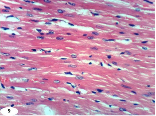
Photomicrographs of heart sections of doxorubicin-injected group treated with quercetin showing an improvement of the kidney. (H and E X 400).
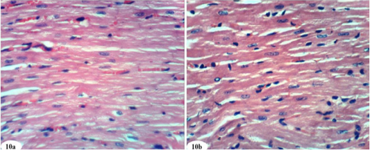
Photomicrographs of heart sections of doxorubicin-injected group treated with mixture of rutin and quercetin showing an improvement of the kidney. (Photomicrographs 10a and 10b). (H and E X 400).


