Advances in Animal and Veterinary Sciences
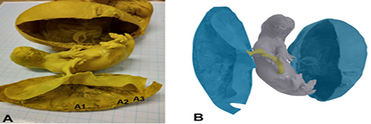
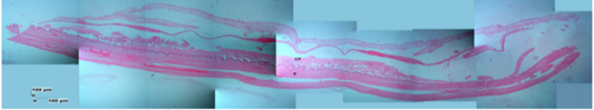
Photomacroscopic and schematic drawing of Sunda porcupine placenta. A is a macroscopic picture of fetal and Sunda porcupine placenta consist of thickes (A1), thin (A2), and thinest (A3) disc parts. B is a schematic drawing of A: blue is a placenta, yellow is umbilical cord, and grey is fetal.
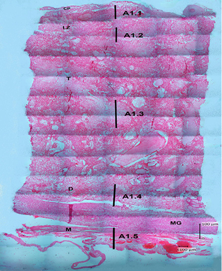
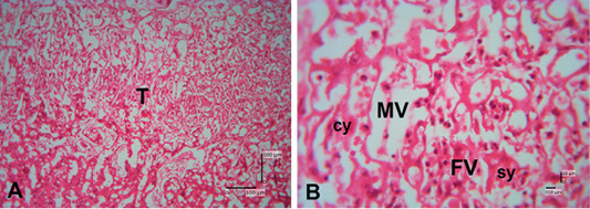
Photomicrograph of the thickest parts of Sunda porcupine placenta refers to Figure 1. A1 (HE). A1.1 is the deepest part, A1.2 is the second part after the innermost part, A1.3 is the middle part, A1.4 is the second outer part, A1.5 is the outermost part of the Sunda porcupine placental thickest disc. chorioallantoic plate (CP), labyrinth zone (LZ), trophospongium (T), decidua (D), metrial glands (MG), myometrium zone (M).
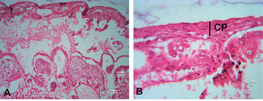
Photomicrograph of chorioallantoic plate area of Sunda porcupine placental disc (A1.1; HE). (A) is the low (125x), (B) are in high magnification (500 x).
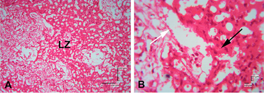
Photomicrograph of labyrinth zone of Sunda porcupine placental disc (A1.2; HE). (A) is in low (125 x) and (B) is in high magnification (500 x). LZ is the labyrinth zone. The white arrow shows the maternal epithelial cells that protrude to form villi. The black arrow shows trophoblast cells in the fetal part of the labyrinth zone.
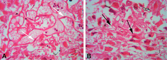
Photomicrograph of trophospongium area of Sunda porcupine placental disc (A1.3; HE). (A) 125x, and (B) 500x magnification. The trophospongium area at low (A) and high magnification (B). At (B) the trophospongium area consist of cytotrophoblast (cy) and syncythiotrophoblast (sy) of the placental margin situated near to the maternal vessel (MV) and fetal vessel (FV).
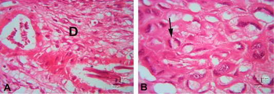
Photomicrograph of decidual area of Sunda porcupine placental disc (A1.4; HE). A and B are in high magnification (500 x). The white arrow shows the maternal epithelial cells that protrude to form villi. The black arrow shows trophoblast cells in the fetal part of the decidua zone.
Photomicrograph of decidual area of Sunda porcupine placental disc (A1.5; HE). The decidua area at low (A) (125x) and high magnification (B) (500 x). At high magnification, decidua cells clearly seen (black arrow).
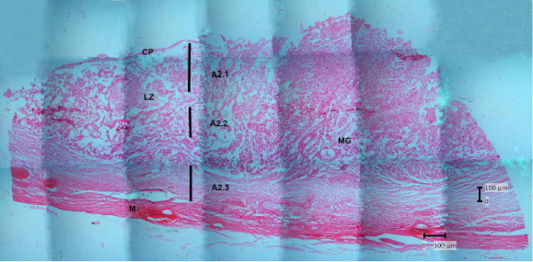
Photomicrograph of the thin parts of Sunda porcupine placental disc refers to Figure 1. A2 (HE). A2.1 is the deepest part, and A2.3 is the outer part. chorioallantoic plate (CP), labyrinth zone (LZ), metrial glands (MG), myometrium (M).
Photomicrograph of the thinest parts of Sunda porcupine placental disc refers to Figure 1. A3 (HE). chorioallantoic plate (CP), endometrium-myometrium layers (E).


