Advances in Animal and Veterinary Sciences
Research Article
Advances in Animal and Veterinary Sciences 1 (4S): 20 – 23Special Issue–4 (Progress in Research on Viruses and Viral Diseases)
A Novel Method of Staining of RNA in Polyacrylamide Gel Electrophoresis
Prasad Minakshi*, Koushlesh Ranjan, Pawan Kumar, Gaya Prasad
-
Department of Animal Biotechnology, Lala Lajpat Rai University of Veterinary and Animal Sciences, Hisar, India
*Corresponding author:minakshi.abt@gmail.com
ARTICLE CITATION:
Minakshi P, Ranjan K, Kumar P and Prasad G (2013). A novel method of staining of RNA in polyacrylamide gel electrophoresis. Adv. Anim. Vet. Sci. 1 (4S):20 – 23.
Received: 2013–10–09, Revised: 2013–11–09, Accepted: 2013–11–10
The electronic version of this article is the complete one and can be found online at
(
http://nexusacademicpublishers.com/table_contents_detail/4/135/html
)
which permits unrestricted use, distribution, and reproduction in any medium, provided the original work is properly cited
ABSTRACT
Polyacrylamide gel electrophoresis (PAGE) is a commonly used technique for analysis of RNA samples because of its low cost, ease of use and high sensitivity. Following one dimensional or two dimensional electrophoresis of RNA sample on a gel, the RNAs are typically visualized by some form of nucleic acid staining techniques. Of these commonly used methods employ use of intercalating fluorescent agents such as ethidium bromide (EB), SYBR green and SYBR gold under short wavelength UV Irradiation. Unfortunately these staining methods are potentially mutagenic and UV radiations are hazardous to both DNA samples and persons handling it. A novel method of RNA staining has been developed using silver ion sensitizer, Eriochrome Black t (EBT). Bluetongue viral RNA resolved in non–denaturing RNA polyacrylamide gel electrophoresis (RNA–PAGE) was used to develop the staining protocol. The method using Eriochrome Black t with silver staining (EBT–SS) has been found to be eight times more sensitive as compared to routine method used in the laboratory. In the routine method of silver staining procedure all the ten bands of bluetongue virus genome were visualized up to 0.625ng RNA sample per lane. However, in newly developed method, using EBT–SS the same was visualized up to 0.078ng RNA sample per lane. The silver ion sensitizer method could be applied for detection of RNA in various biological samples due to its higher sensitivity.
INTRODUCTION
Polyacrylamide gel electrophoresis (PAGE) is a commonly used technique for analysis of RNA samples because of its low cost, ease of use, and high sensitivity. Following one–dimensional or two–dimensional electrophoresis of RNA samples on a gel, the RNAs are typically visualized by some form of nucleic acid staining techniques. Of these, the most popular techniques in use are intercalating fluorescent agents, such as ethidium bromide (EB), SYBR green, and SYBR gold, under short–wavelength UV irradiation. Unfortunately, these staining methods are potentially mutagenic and UV irradiations are hazardous to both the DNA sample and persons handling it.
The silver staining technique was originally standardized for staining of protein gels (Switzer et al. 1979). Later on the method was used for nucleic acid staining (Blum et al., 1987; Goldman and Merril, 1982). Currently, silver staining has been widely been used for detection of nucleic acid (DNA and RNA fragments) in various experiments. The sensitivity of silver staining is the comparable to radio isotopic methods. However, the conventional Silver staining method is complicated and time–consuming because they need a lot of preparation and handling of several solutions prior to use. Moreover, the colour uniformity, sensitivity and storage time of the stained gels are not much ideal with conventional silver staining method.
Radio labelling of nucleic acid with radioactive isotopes is likely to remain the most sensitive method available. However, this method is hazardous and requires a complicated handling procedure that limits its application. The silver staining methods as described in literature have an advantage in term of sensitivity compared to EB staining, but the sensitivity of silver staining still remain at a level of 0.01–1 ng/band (Creste et al., 2001). The sensitivity of silver staining of PAGE for DNA can be increased dramatically using a silver ion sensitizer such as Eriochrome black–t (EBT) along with silver ion (Hwang et al., 2006; Han et al., 2008). The silver staining along with EBT had been used for sensitive detection of protein in SDS–PAGE. The detection limit was found to be 0.05–0.2 ng protein within 60 min in SDS–PAGE gels (Jin et al., 2006). However, the EBT–silver staining for RNA–PAGE has not been described to best of our knowledge.
In the present study, a standard method of silver staining routinely used in our laboratory (Svensson, et al., 1986) for RNA–PAGE staining is compared with an ultrasensitive EBT–silver staining for RNA staining in non–denaturing polyacrylamide gel originally described for staining of DNA in PAGE (Hwang et al., 2006).
MATERIALS AND METHODS
Virus Sample Cultivation
The BTV serotype 16 was cultivated in BHK–21 cell line using minimum essential medium (Sigma, USA). After appearance of 75% of cytopathetic effect (CPE) the virus was harvested.
Extraction of Viral Nucleic Acid
The cell culture grown virus sample was allowed to centrifugation at 8000 rpm (Remi, India) to pellet down the BHK21 cells along with virus. The viral nucleic acid (dsRNA) was extracted from pelleted material using Tri reagent (Sigma, USA) as per manufacturer’s instruction. Two fold serial dilutions of viral dsRNA were done to analyse in RNA–PAGE.
RNA Polyacrylamide Gel Electrophoresis (RNA–PAGE)
Two sets of eight percent non–denaturing polyacrylamide gels (80 mm X 100mm X 1.0 mm) were prepared as per standard protocol (Sambrook and Russell, 2001). In Brief, 30% Acrylamide/Bisacrylamide mixture (29.2g Acrylamide: 0.8 g Bisacrylamide), 1.5 M tris HCl pH 8.8, Ammonium per sulphate (10%), N' N' N' N' tetra methylene ethylene diamine (TEMED) and glass distill water were mixed homogenously and poured between glass plates. All the reagents used are of A.R. grade manufactured from SRL, India. The two fold serially diluted viral nucleic acids were loaded onto the two sets of gel having 5, 2.5, 1.25, 0.625, 0.312, 0.156, 0.078, 0.039, 0.019 ng of dsRNA per lanes. Electrophoresis was carried out using discontinuous buffer system (Laemmli, 1970) in gel electrophoresis system (BioRad, USA), using power supply at 12V/cm and Tris–Glycine was used as the running buffer.
Staining of Polyacrylamide Gel
Silver Staining of Gel
The gel was removed from electrophoresis assembly and fixed in100ml of 10% ethanol (Merck, India) and 0.5% glacial acetic acid (SRL, India) at room temperature for 30 minute with gentle shacking. Subsequently the gel was stained in 100ml of 0.185% Silver nitrate (SRL, India) for 30 minute with gentle shacking. Extra silver stain was removed by quick washing with glass distil water. dsRNA bands were visualized using 100ml of developer (3% NaOH and 0.75% formaldehyde) for 2–3 minute. Reaction was stopped with 5% glacial acetic acid and finally stored in 10% ethanol (Table 1).

Table 1: Comparative protocol of routine silver staining and EBT–Silver staining for RNA in polyacrylamide gel
EBT–Silver staining (EBT–Silver Staining)
After electrophoresis, another set of gel was fixed in 100 mL of 40% ethanol containing 10% glacial acetic acid for 20 minutes two times. The gel was sensitized for 1 minute with 50 mL of 0.0025% EBT (SRL, India) in glass distil water. It was destained by 100 mL of fixing solution for 1 min, and finally in 100 mL of glass distilled water for 2 minutes twice. The gel was subjected to 50ml of 0.25% silver nitrate containing 0.037% formaldehyde solution for 5 minute at room temperature. The gel was washed with 100 mL of glass distilled water for 10 second twice, and then reduced in 50 mL of 2% potassium carbonate solution containing 0.01 M sodium hydroxide, 0.014% formaldehyde and 0.0002% sodium thiosulfate for 3–5 minute. Finally, the gel was immersed in 50 mL of 1.5% EDTA for 10 min and washed with 50 mL of glass distil water for 5 min to stop the reaction. The gel was stored in 10% ethanol (Table 1).
RESULTS
The viral dsRNA from BHK–21 cell culture grown was extracted in our lab. The dsRNA was serially two fold diluted from 5ng/µl to 0.019ng/µl and sequentially loaded to the two sets of non–denaturing polyacrylamide gels. The one of the polyacrylamide gel was stained with routine silver staining whereas the other gel was allowed for EBT–Silver staining (Table 1). The gels photograph were recorded by a photo camera (Nikon, Japan) using a trans–illuminator (Figures 1 and 2). The sensitivity of routine silver staining result was compared with that of EBT–Silver staining. In routine silver staining procedure all the ten bands of bluetongue virus genome were visualized up to lane 4 having 0.625 ng of dsRNA sample. In contrast to the routine silver staining method in EBT–Silver staining procedure the viral dsRNA was visualized up to lane 7 having 0.078 ng RNA sample.
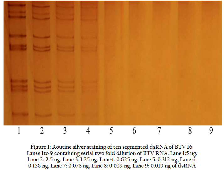
Figure 1: Routine silver staining of ten segmented dsRNA of BTV 16. Lanes 1to 9 containing serial two fold dilution of BTV RNA
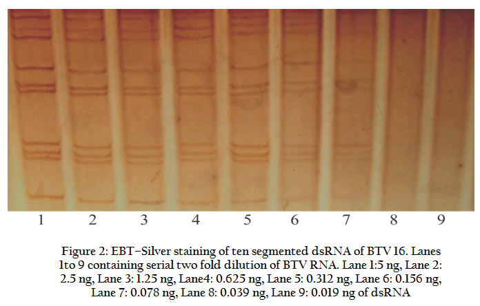
Figure 2: EBT–Silver staining of ten segmented dsRNA of BTV 16. Lanes 1to 9 containing serial two fold dilution of BTV RNA
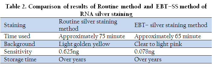
The sensitivity, total staining time taken etc. of both the methods were compared (Table 2). It showed that the detection limit of EBT–Silver staining method for dsRNA is approximately 8 folds higher than that of the routine silver staining method. Moreover, EBT–silver staining method takes approximately 10 minutes less time in entire staining procedure.
DISCUSSION
The RNA–PAGE along with silver staining is a commonly used technique for detection of viral nucleic acid because of its known sensitivity and relatively less time taking procedure. In general silver staining method of RNA is relatively sensitive. However, one of the most efficient ways to increase sensitivity is the addition of an enhancing step prior to the impregnation by silver ions. In Present study, EBT dye was used as an efficient silver–ion sensitizer for RNA silver staining. Although, the mechanism by which EBT–SS works is not yet clearly understood however, the diazo bond of EBT has the ability to reduce silver ions in alkaline solutions. Moreover, silver ions can be reduced further by dihydroxynaphthalenes produced from EBT cleavage. Probably these properties of EBT increase the sensitivity and speed of EBT–Silver staining.
Fixation is the first step for gel staining after electrophoresis which provides RNA binding to the gel matrix and allow for the removal of the buffer components from gels. Reductants such as formaldehyde are added to the silver nitrate solution directly to facilitate the deposition of silver ions in RNA zones. Therefore, in the present method, 0.037% formaldehyde was used with silver nitrate in the impregnation step. In addition, since the EBT has a reducing power itself, formaldehyde would have an enhanced reducing effect with the EBT. However, in routine silver staining method, staining solution can be reused for 2–3 times if properly stored under dark conditions. The same can not be reused in EBT–SS method because the silver solution is premixed with formaldehyde, hence slightly increasing the cost of the test. Moreover, for better result in EBT–Silver staining method freshly prepared 0.0025% EBT solution is recommended.
The EBT silver staining method is superior to the routine silver staining method in terms of time used in entire procedure, sensitivity, colour uniformity and background development. The routine silver staining method used to take approximately 75 minute in entire staining procedure. However, the EBT–silver staining method takes slightly less approximately 65 minute. The background colour of gel of routine silver staining method was light golden yellow. Whereas, the EBT–silver staining showed clear to light pink background colour, which provided better visibility of nucleic acid (dsRNA) bands in gel. The gel stained with both of these methods described can be stored in 10% ethanol for over a year without change in intensity of nucleic acid (dsRNA) bands and breakage of gel.
The similar EBT–silver staining method was described for DNA. The minimum detection limit was found to be 1 pg for φX174 DNA/HaeIII in both non–denaturing and denaturing polyacrylamide gels (Hwang et al., 2006). In one another assay using EBT–silver staining the detection limit was found to be the minimum of 1.75 ng of DNA of pBR322 DNA/MspI DNA marker sample in non–denaturing polyacrylamide gel (Han et al., 2008). The above described assays were standardised using the known commercially available DNA marker. To the best of the knowledge of the authors, there is no report on the application of EBT for staining of RNA by RNA–PAGE in the literature. This could probably be the first report of detection of RNA from biological sample by using EBT–Silver staining in RNA–PAGE.
CONCLUSIONS
To summarize the findings of the present study, the novel method developed for detection of viral RNA by RNA–PAGE using EBT–Silver staining has been found 8 times more sensitive as compared to routinely used RNA–PAGE–Silver staining. Further, the EBT–silver staining method described could be useful for detection of RNA of other viruses in cell culture supernatants such as rotavirus as well as RNAs isolated from various biological samples such as blood, semen, tissues and faecal samples due to its higher sensitivity.
ACKNOWLEDGMENTS
Authors are thankful to AINP–BT, ICAR, New Delhi and Department of Animal Biotechnology, LLR University of Veterinary and Animal Sciences, Hisar for providing funds and sufficient infrastructure respectively to carry out the study.
CONFLICT OF INTEREST
We declare that we don’t have any conflict of interest.
REFERENCES
Blum H, Beier H and Gross HJ (1987). Improved silver staining of plant proteins, RNA and DNA in polyacrylamide gels. Electrophoresis. 8:93 – 99.
http://dx.doi.org/10.1002/elps.1150080203
Creste S, Tulmann NA and Figueira A (2001). Detection of Single Sequence Repeat Polymorphisms in Denaturing Polyacrylamide Sequencing Gels by Silver Staining. Plant Mol Biol Rep.19:299 – 306.
http://dx.doi.org/10.1007/BF02772828
Goldman D and Merril CR (1982). Silver staining of DNA in polyacrylamide gels: Linearity and effect of fragment size. Electrophoresis. 3:24 – 26.
http://dx.doi.org/10.1002/elps.1150030105
Han YC, Teng CZ, Hu ZL and Song YC (2008). An optimal method of DNA silver staining in polyacrylamide gels. Electrophoresis. 29:1355 – 1358.
http://dx.doi.org/10.1002/elps.200700558
PMid:18348333
Hwang SY, Jin LT, Yoo GS and Choi JK (2006). Silver staining method for DNA in polyacrylamide gels using eriochrome black T as a silver–ion sensitizer. Electrophoresis. 27:1744 – 1748.
http://dx.doi.org/10.1002/elps.200500601
PMid:16568502
Jin LT, Hwang SY, Yoo GS and Choi JK (2006). A mass spectrometry compatible silver staining method for protein incorporating a new silver sensitizer in sodium dodecyl sulfate–polyacrylamide electrophoresis gels. Proteomics. 6:2334 – 2337.
http://dx.doi.org/10.1002/pmic.200500596
PMid:16493709
Laemmli UK. (1970). Cleavage of structural proteins during the assembly of head of bacteriophage T4. Nature. 227:680 – 685.
http://dx.doi.org/10.1038/227680a0
PMid:5432063
Sambrook JD, Russell WA and Jassen K (2001). Molecular cloning: a laboratory manual. 3rd edn. Cold Spring Harbour Laboratory Press, New York.
Svensson L, Uhnoo I, Grandien M and Wadeli G (1986). Molecular epidemiology of rotavirus infections in Upsala. Sweden. 1981; disappearance of a predominant electropherotype. J Med Virol. 18:101 – 111.
http://dx.doi.org/10.1002/jmv.1890180202
PMid:3005484
Switzer Rc 3rd, Merril CR and Shifrin S (1979). A highly sensitive silver stain for detecting proteins and peptides in polyacrylamide gels. Anal Biochem. 98:231 – 237.
http://dx.doi.org/10.1016/0003-2697(79)90732-2


