Advances in Animal and Veterinary Sciences
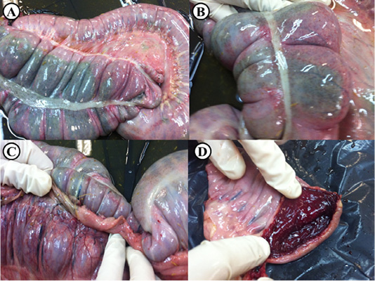
Post-mortem lesions of an adult horse suspected to be infected with C. difficile. (A) Diffuse darker discoloration of the serosal surface of the colon, (B) Sub-serosal petechiae are seen on the affected colon, (C) Colonic wall blood vessels are severely congested, (D) Opened colon shows marked congestion and hemorrhage of the mucosal surface.
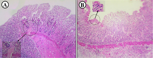
Histopathological findings in an adult horse suspected to be infected with C. difficile. (A) Colon showing coagulative necrosis of the mucosa and prominent mucosal and submucosal thrombosis (inset), (B) Colon showing congestion and necrosis in addition to diffuse infiltration of leucocytes and fibrinous exudate (inset).
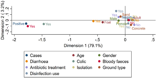
Multiple correspondence analysis of risk factors associated with the presence of C. difficile toxins in diarrheic and normal adult horses and foals.


