Advances in Animal and Veterinary Sciences
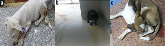
Puppies suffering from CPV infection, a. Weakness, dullness, and diarrhea. b. Weakness, dullness, vomiting and bloody diarrhea. c. Weakness, dullness and bloody diarrhea
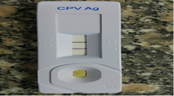
CPV-positive sample by rapid Chromatographic slide immunoassay.
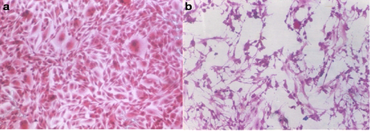
a. Normal Vero cell culture. b. Vero cell culture infected with the obtained CPV showing distorted cell morphology and detached cells (H&E; 100Xs)
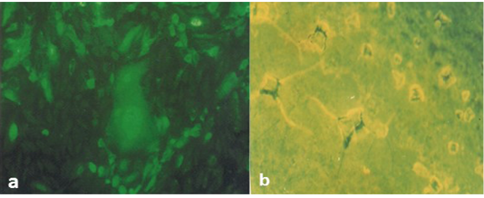
(a) Positive FAT showing intra-cytoplasmic apple green reaction. (b) Negative FAT.
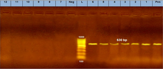
PCR assay using CPV-2 primers. (L) DNA ladder 100 bp, lanes (1: 6) positive samples and specific bands at 630 bp. Lane (Pos) : positive control. Lanes (7:12) negative samples, lane (neg) negative control sample.
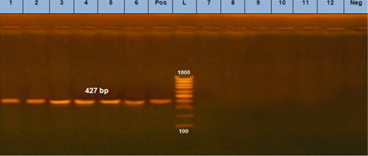
PCR assay using using primer set CPV- 2a. (L) DNA ladder 100 bp, lanes (1: 6) positive samples and specific bands at 427 bp. Lane (Pos): positive control. Lanes (7:12) negative samples, lane (neg) negative control sample.
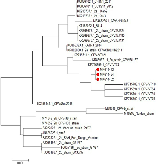
Phylogenetic tree of CPV isolates in this study compared to other sequences in the gene bank.


