Advances in Animal and Veterinary Sciences
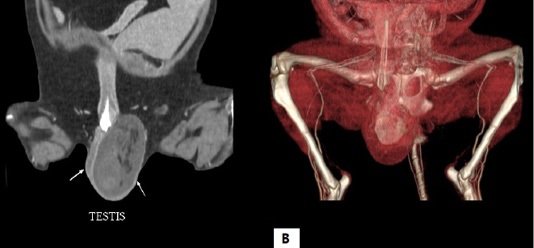
Computed tomography scan of the testicle. (A) Two-dimensional computed tomography scan shows hyperdensity of left testicle and (B) three-dimensional computed tomography scan presents a mass within the left testicle.
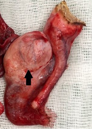
Gross lesion of the surgically removed tissue. A neoplastic mass with a diameter of 1 centimeter was observed in the tunica vaginalis (arrow).
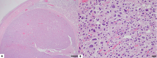
Histopathological lesion of the tumor. (A) An encapsulated neoplastic mass has proliferated and compresed adjacent atrophic seminiferous tubules (H&E stain, Bar = 1mm). (B) Neoplastic cells showed marked nuclear pleomorphism, anisokaryosis, and distinct single nucleolus (H&E stain, Bar = 20 µm).
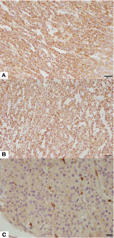
Immunohistochemistry for definite diagnosis of the Leydig cell tumor. Neoplastic Leydig cells showed homogenous positivity for (A) vimentin (Counterstained with Mayer’s hematoxylin, Bar = 50 µm), (B) Melan A (Counterstained with Mayer’s hematoxylin, Bar = 50 µm), and (C) c-kit (CD117) (Counterstained with Mayer’s hematoxylin, Bar = 20 µm).


