Advances in Animal and Veterinary Sciences
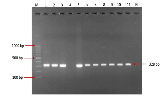
Gel electrophoresis image showing: Lane M) Exact Mark 100 – 1000 bp DNA ladder, Lane 1-11 except 4) conventional PCR technique detected all possible FMDV in approximately band size 328bp. Lane 4) is negative result, Lane N) DNA extracted from FMDV-free cattle used as negative control.
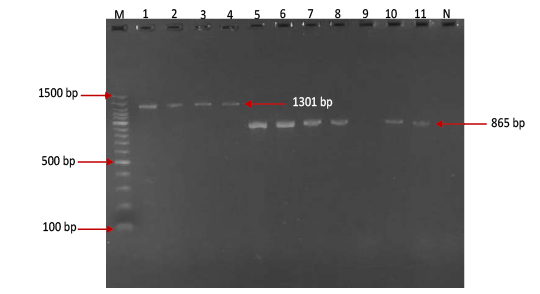
Gel electrophoresis image showing Lane M) Exact Mark 100 – 1500 bp DNA ladder, Lane 1-4) conventional PCR technique detecting genotype O of FMDV in approximately band size 1301 bp. Lane 5-11except 9) conventional PCR technique detecting genotype A of FMDV in approximately band size 865 bp, Lane 9) is negative result, Lane N) DNA extracted from FMDV-free cattle used as negative control.
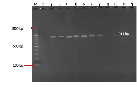
Gel electrophoresis image showing Lane M ) Exact Mark 100 – 1500 bp DNA ladder, Lane 2-8) conventional PCR technique detecting genotype Asia-1 of FMD virus in approximately band size 911 bp. Lane1,9,10,11) are negative results, Lane N) DNA extracted from FMDV-free cattle used as negative control.
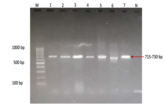
Gel electrophoresis image showing Lane M) Exact Mark 100 – 1000 bp DNA ladder, Lane 1-7) conventional PCR technique detecting only genotype SAT of FMD virus in approximately band size 715- 730 bp. Lane N) DNA extracted from FMDV-free cattle used as negative control.


