Advances in Animal and Veterinary Sciences
Research Article
Adv. Anim. Vet. Sci. 7(12): 1134-1139
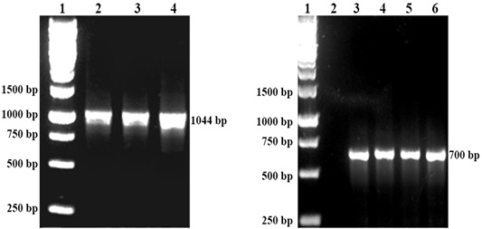
Figure 1
Left: DNA detection of P. multocida by PCR. Lane 1: Protein marker, Lane 2: P. multocida A:1, Lane 3: P. multocida A:3 Lane 4: P. multocida A:1,3. Right: DNA detection of R. anatipestifer by PCR. Lane 1 Protein marker. Lane 3,4,5 and 6: R. anatipestifer.
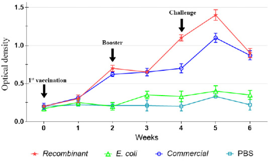
Figure 2
IgG antibody against P. multocida A:1 response in chickens during the immunization and challenge trial.
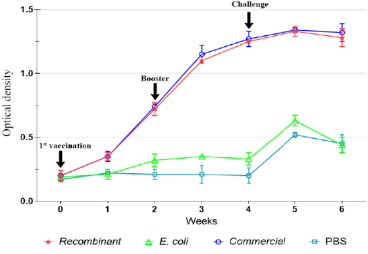
Figure 3
IgG antibody against P. multocida A:1 response in ducks during the immunization and challenge trial.
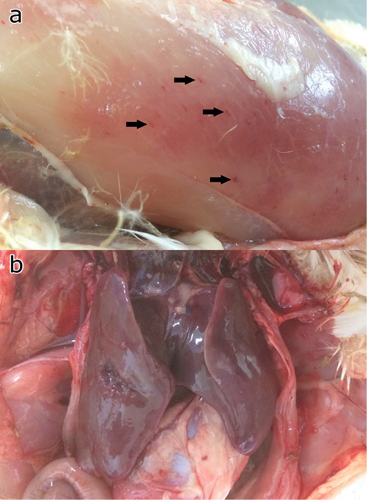
Figure 4
Gross pathology of the chicken and ducks showing petechia haemorrhage (arrows) of the pectoral muscle (a), and hepatic congestion (b).


