Advances in Animal and Veterinary Sciences
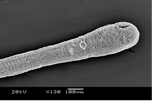
Scanning electron microscopy of the anterior portion of R. galli treated with M. taiwaniana extract. The terminal bulb is the scolex and the stalk is the neck. Two suckers are seen on the scolex. x 130, scale bar is 100 µm.
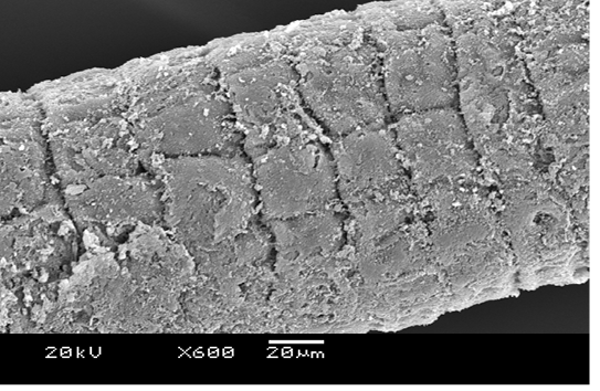
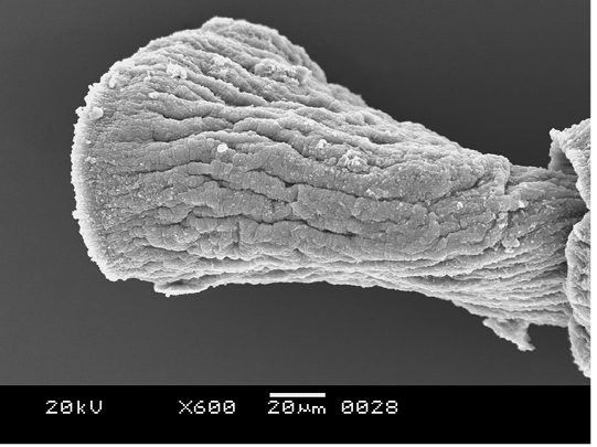
Scanning electron microscopy of the neck of R. galli treated with M. taiwaniana extract. Body segments or proglottids are heavily eroded. x 600, scale bar is 20 µm.
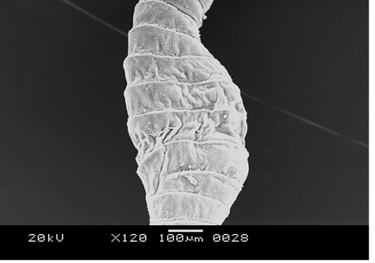
Scanning electron microscopy of mature proglottids of R. galli treated with M. taiwaniana extract. Abnormal enlargement of the body is evident. x 120, scale bar is 100 µm.
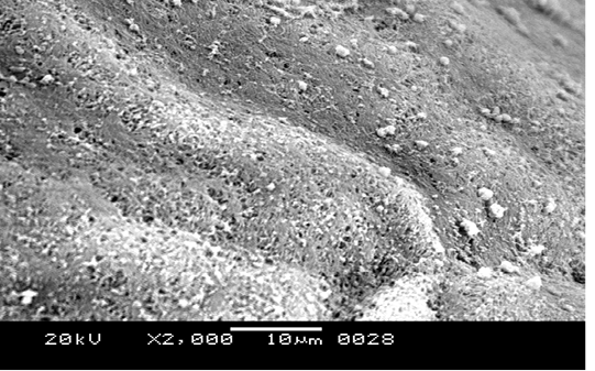
Scanning electron microscopy of a mature proglottids of R. galli treated with M. taiwaniana extract. A fuzzy surface depicts degenerated microtriches. x 2000, scale bar is 10 µm.
The last gravid proglottid of R. galli treated with M. taiwaniana extract. Shrinkage with a series of longitudinal folds is visible. x 600, scale bar is 20 µm.
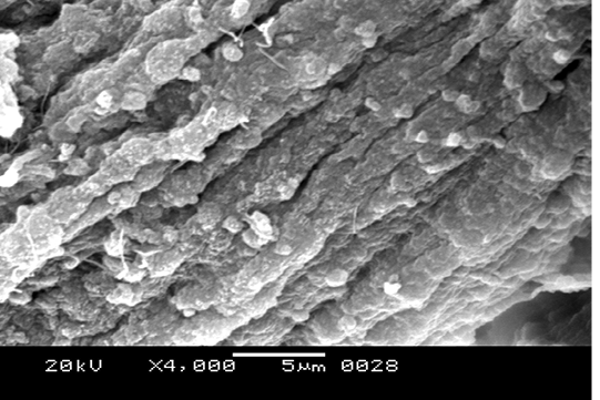
Magnification of the tegument of a gravid proglottid of R. galli treated with M. taiwaniana extract. Tegumental folds are composed of blisters or blebs. x 4000, scale bar is 5 µm.


