Advances in Animal and Veterinary Sciences
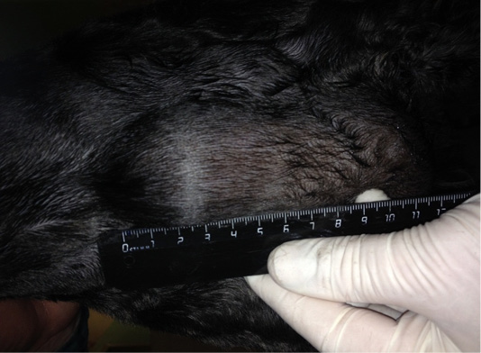
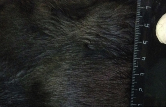
Neoplasm of the mammary gland in the M2 region on the right side. Doggess, miniature schnauzer breed, at the age of 10 years.
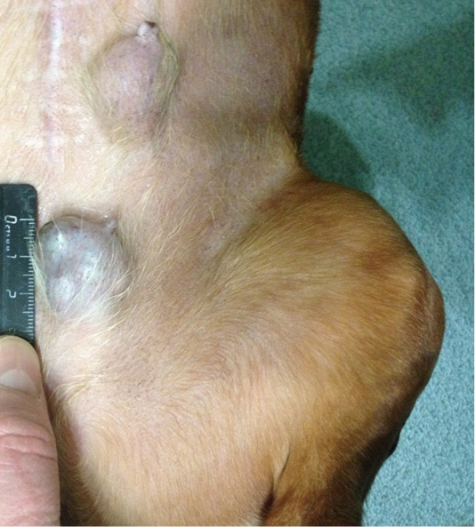
Localization of tumor nodules: M4 and M5 region on the left side. Doggess, breed dachshund standard, at the age of 10.5 years.

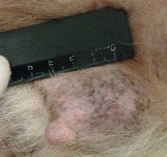
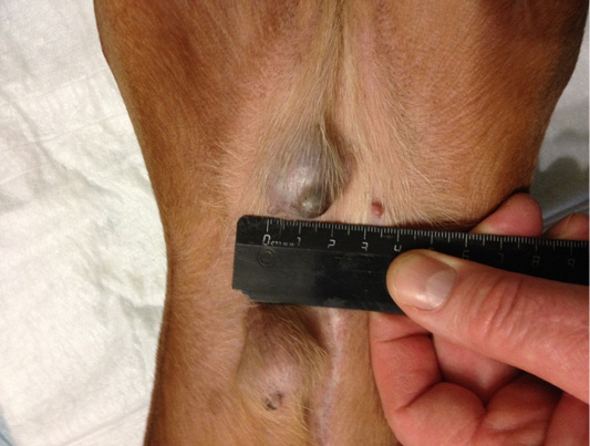
Mammary gland neoplasm in the M1 region on the right side. Doggess, breed jagdterrier, at the age of 12 years.
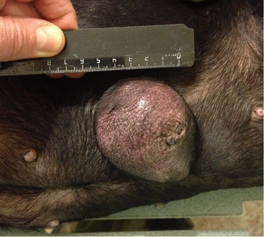
Mammary gland neoplasm in the M4 region on the left side. Doggess, American Bulldog, at the age of 11.5 years.
Metastasis of breast cancer in the liver parenchyma. Doggess, breed American Bulldog, at the age of 11.5 years.
Localization of the tumor in M4 region on the left side. Doggess, Miniature Schnauzer, at the age of 10.5 years.
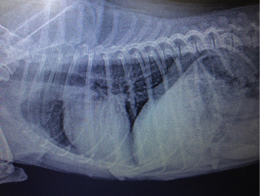
Metastasis of mammary gland cancer in the lungs. Doggess, Miniature Schnauzer, at the age of 10.5 years.
Localization of tumor nodules in M4 and M5 regions on the right side. Doggess, dachshund standard, at the age of 9 years.
Localization of the tumor site M2 on the right, in the area of the mastectomy previously performed. Doggess, standard dachshund breed, age of 11.5 years.
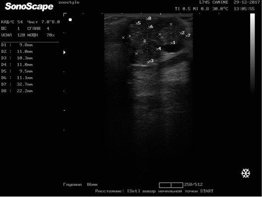
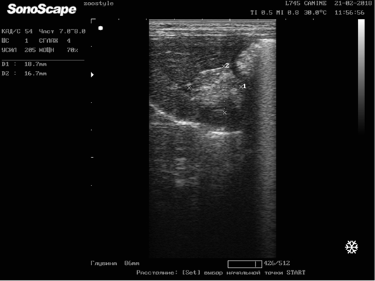
Longitudinal scanning of the mammary gland tumor (M5 region on the right side) on day 42 of the therapy (32.7х22.2 mm). Doggess, American Bulldog, at the age of 11.5 years.


