Advances in Animal and Veterinary Sciences
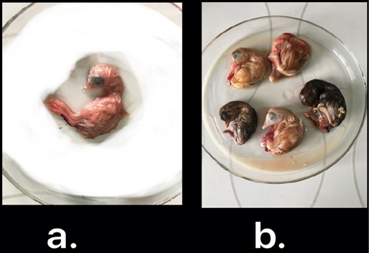
IBV isolated in ECEs. Normal embryo development (a) and curled, stunted, and dwarfed embryos infected with IBV after fourth passage (b)
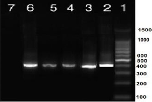
RT-PCR amplification of partial S1 gene from IBV isolates in the present study showed a single specific band (400bp). Lane 1: 1 kbp DNA ladder (Fermantas); Lane 2, positive control; Lanes 3–6: positive IBV-infected samples. Lane 7: negative control.
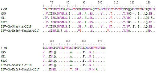
Comparison between deduced amino acid sequences of HVR1 (amino acid position from 60-88) and HVR2 (amino acid position from 115-140) of S1 protein of two isolates reported in the study and currently used vaccine strains in Egypt.
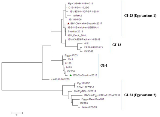
Phylogenetic tree based on nucleotide sequences of S1 gene for the two IBV isolates and related 25 reference IBV strains. The IBV-Ch-KafrAshaykh-2017 isolate marked with a red triangle belonging to isolates of GI-23 lineage (Egy/variant1 group), while IBV Ch-Sharkia-2018 marked with a green triangle belonging to isolates of GI-1lineage (classical group).


