Advances in Animal and Veterinary Sciences
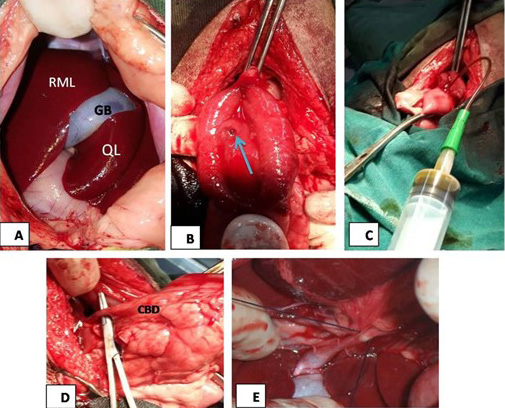
(A) The mid line incision at the epigastric region in liver area, RML: right medial lobe of liver, GB: gallbladder, QL: quadrate lobe; (B): image showed duodenal incision and exposure of major duodenal papilla (arrow); (C): the image showed bile aspiration; (D): the image showed common bile duct (CBD) exposure; (E): surgical ligation of common bile duct.
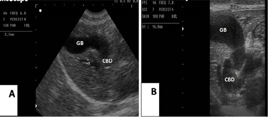
(A): Sagittal ultrasonographic image of gallbladder (GB) and common bile duct (CBD) in dog before surgery, 2.7mm; (B): the image showed the maximum dilatation of gallbladder (GB) and common bile duct in the 2nd week after ligation, 14.9mm.
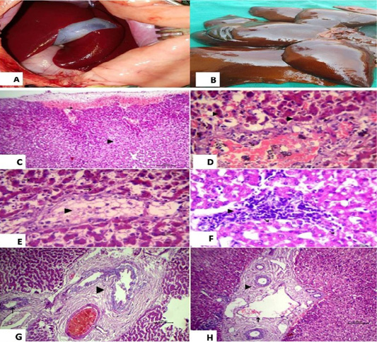
(A): showed normal liver before surgery; (B): Liver showed necrosis and firm in consistency after ligation and showed multiple grayish necrotic areas. Photomicrograph of HandE stained liver tissue; (C): subcapsular hemorrhage (arrow) and vacuolation in some hepatocytes (arrow head), 100 bar; (D): congestion of central vein (black arrow), vacuolation in some hepatocytes (white arrow) and yellowish brown granules of bile pigment inside other hepatocytes (arrow head), 20 bar; (E): fatty change of some hepatocytes (arrow head) and disassociation of other hepatocytes (white arrow) with yellowish brown granules of bile pigment inside hepatocytes HandE 20 bar; (F): the focal aggregation of lymphocytes with few macrophages infiltrations replaced necrotic hepatocytes (arrow head) and vacuolation in other hepatocytes, 20 bar; (G) Chronic cholecystitis represented by hyperplasia of its lining epithelium and thickening in the wall of gall bladder with fibrous tissue proliferation (arrow head) and chronic cholangitis (arrow), 100 bar; (H) Dilation of portal vein (black arrow) with chronic cholangitis represented by thickening in the wall of bile duct with fibrous tissue proliferation (arrow head), 100 bar.


