Advances in Animal and Veterinary Sciences
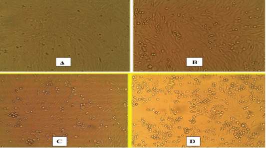
Microscopic images of inoculation, propagation and isolation of PPRV on Vero cells for 3 successive blind passages; (A) Non-inoculated monolayer Vero cells (Control) recorded at Zero day show confluent monolayer sheath; (B) Vero cells inoculated with PPRV suspected sample for 1st blind passage with CPE recorded at 6th dpi represented by primary cell rounding; (C) Vero cell line at 2nd passage inoculated blindly with supernatant harvest of 1st passage with development of CPE at 5th dpi showing more rounding, detachment and gapping of the cells; (D) Vero cells inoculated at 3rd blind passage level with clear and marked CPE include complete rounding, cluster aggregations appearance of the cells with detachment. Gapping of the cells in between.
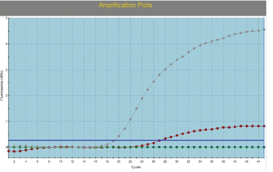
Real time Reverse Transcription Polymerase Chain reaction (RT-qPCR) for detection of PPRV in infected Vero cells.


