Advances in Animal and Veterinary Sciences
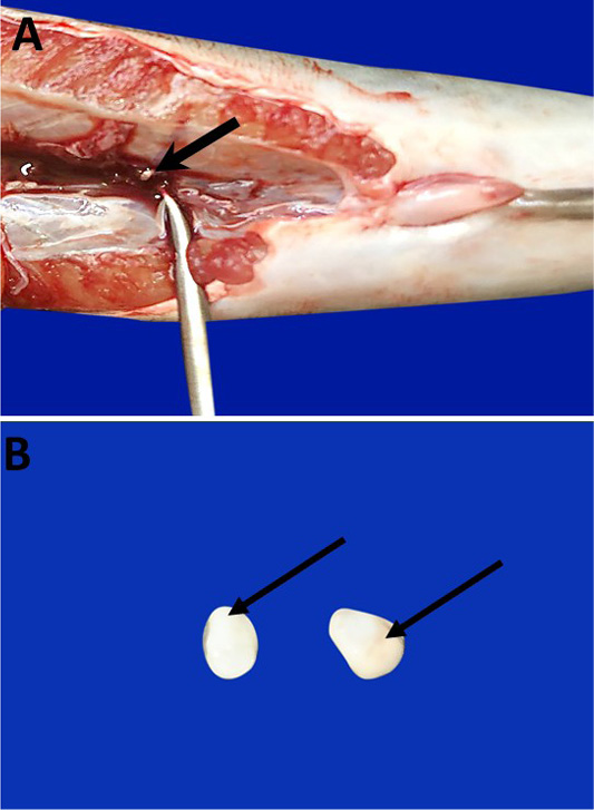
Photomacrographs show Catfish SC which are partially embedded in the posterior region of trunk kidney (bold arrow) (A). They are paired of small, oval, colored white or creamy bodies (arrows) (B).
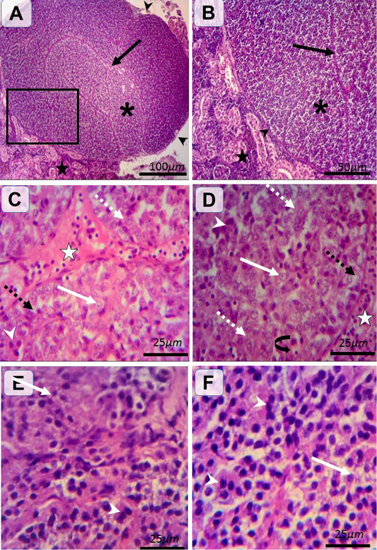
Histological structure of female and male catfishes SC. Representative photomicrographs of HandE stained sections illustrating renal tissues (black stars), C.T. capsule (black arrow heads), septa (black arrows), SC of incomplete lobules (asterisk) (A and B). Type I (weakly stained) cells are rounded-shaped with pale homogenous eosinophilic (white dashed arrows) or vacuolated (black dashed arrows) cytoplasm and lightly stained basophilic rounded nuclei with prominent nucleoli (white arrows). Type II (intensely stained) cells are irregular-shaped with deeply eosinophilic cytoplasm and darkly stained basophilic rounded nuclei (white arrow heads) (C, D, E andF). The pronounced cells are type I in female (C andD) and type II in male (E andF). Notice the C.T. septa housing blood vessels (white star), plentiful blood capillaries within gland (closed arrow).
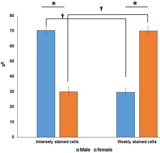
Chart showing (*) significant differences in the ratio of the two cell types in the same sex. Furthermore, (†) significant differences are observed between the two types of cells in between both sexes.
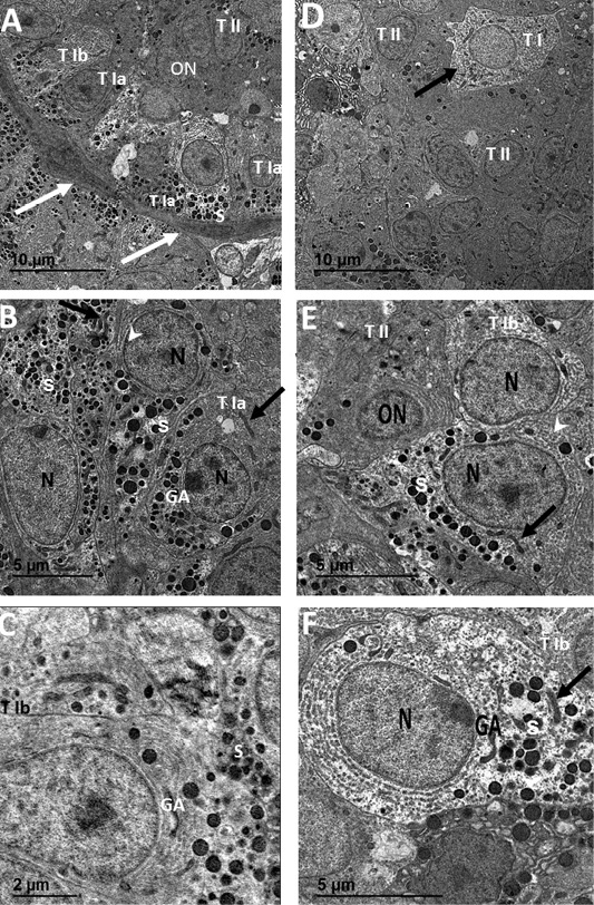
Transmission electron micrographs of SC from female catfish (A, B and C) showing septa housing numerous blood capillaries (white arrows) and surrounding follicular glandular cells of Type I with two subtypes; Type Ia (T1a), Type Ib (T1b) and Type II (TII), Type I cell with concentrated secretory granules (S) near capillaries and rER (arrowhead) on the opposite pole, Type II cells with small oval nuclei (ON) of dense heterochromatin patches and electron dense cytoplasm (white arrow) (A). Type Ia (TI a) has electron dense cytoplasm with rounded euchromatic nuclei (N), numerous secretory granules (S), Golgi apparatus (GA) and filamentous mitochondria (black arrows) (B). Type Ib (TI b) has electron lucent cytoplasm with rounded euchromatic nuclei (N), few secretory granules (S), Golgi apparatus (GA) and rER (black arrow head) (C). Transmission electron micrographs of SC from male catfish (D, E and F) showing glandular cells cluster of mainly Type II (TII) and sparse of Type I (T I) cells (A). Type II (TII) has electron dense cytoplasm with small oval nuclei (ON) of dense heterochromatin patches, rER and few secretory granules (S) (B). Type Ib (T Ib) cells have electron lucent cytoplasm with rounded euchromatic nuclei (N), few secretory granules (S), rER (white arrowhead), Golgi apparatus (GA) and filamentous mitochondria (black arrow) (B and C).


